Tuesday, July 31, 2012


Malignant Melanoma

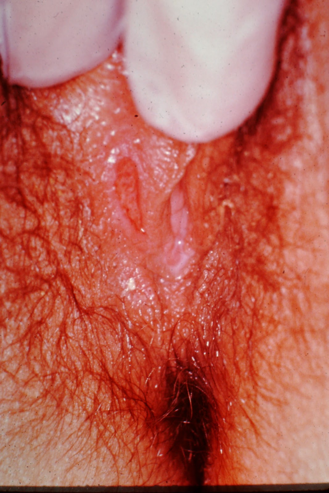
What do you see when you see a lesion such as this?  Do you see merely a melanoma, or do you see more?  By examining this picture, I hope you will start to look at lesions such as this more critically, because your perception dictates your actions, which in turn dictates your outcomes.  So, let us take a "Zen" look at this lesion, and decide what to do.

This lesion is on the trunk of a Caucasian gentleman, and could not more clearly violate all of the ABCDs.  It is asymmetric, irregularly bordered, wildly variegated in coloring, and if one compares the lesion to the hair, is well north of 6 mm in diameter.  

What about the ddx?  My ddx would be a Pigmented SBCC, Pigmented Bowens, and SK.  All three are dismissed easily, though, because of the appearance.  Pigmented Bowen's is more uniform in color, and is usually on darker skin individuals.  Pigmented BCCs would not have the elevation, irregular pigmentation, and would have an accentuated border.  SKs have a different texture, different coloration and, even when traumatized, would not have the same notched border and color variegation of this one.

So, having settled on MM being the likely dx, we choose to do an excisional biopsy.  Why excisional?  Because one never knows which day a melanoma will decide to metastasize.  If you are sure it is MM, cut the damn thing out. Seeing it is large, my preference is as follows:  Excise with narrow, but complete margins because if it is large or shows signs of regression (more on this in a minute) then one can reasonably demand a sentinel node biopsy down the road.  

If the lesion is on an extremity, close the incision, if possible, in the direction of nodal drainage, i.e. longitudinally.  On the trunk, close it via the long axis, to make the overall incision as short as possible. Do not extensively reduce the standing cones (dog ears).  It will theoretically confuse the determination of which is the best nodal basin to sample on SLNB.

Now, looking at the lesion, it is elevated in spots, but not too significantly so.  My estimation was at first 1.5 mm Breslow's but I changed it after a little reflection to 1.1 mm.  It turned out to be .95.  Next, I looked for signs of inflammation.  I saw not only the pink spot, but also a faint pink area extending away from the lesion at 9 o'clock.  Also, I saw two unequivocal signs of regression:  the notching at 12 o'clock and the linear scarring within the lesion.  What this tells me is this:  regardless of whether or not he has palpable lymphadenopathy, his immune system has "presented" the tumor cells to the draining node or nodes, and as a result has at least micrometastasis.  As the late dermatopathologist Bernie Ackerman said, signs of regression are uniformly bad news for the patient.  

Finally, the size of the superficial spreading melanoma tells the tragic part of the story:  It has been there for a very long time.  It was on a visible part of the trunk, on a well-educated man, and he watched it grow for years.  Even when I raised the possibility that it might be melanoma, he seemed surprised, but relatively unconcerned.  He didn't really grasp the gravity of the diagnosis.  How this can be is a mystery to me, but he was blissfully ignorant of the concept of how a lesion such as this could change his world.

This is the thing that all of you live, eat and breathe to fight against.  There is no more worthy goal than for all of you to educate, both in your clinic and in your community, those who, like this man, those who don't know how horrible this disease is.  And maybe, one day, someone will come up to you at the end of a lecture at a church or garden club or civic center, and they will ask you about a small brown spot with irregular borders, and you will tell them to get it biopsied, and they will live long, happy lives because you educated them, and saved their life.  Isn't that a goal worth having?


Monday, July 30, 2012


Sarcoidosis

Sarcoidosis is a systemic disease that is manifested by granulomatous infiltration of multiple organs sites, with the most common being the lungs, followed by the lymph nodes.  Other sites of involvement include the liver, skin and eyes, with the heart, CNS and kidneys bringing up the rear.

The most common way someone is diagnosed with sarcoidosis is via a chest x-ray, followed by physical exam.  Salivary gland enlargement and tear duct changes are physical findings that can clue you in to the diagnosis.  

On the skin, the manifestations are so protean that some people call it the "great imitator", much like they used to call syphilis the same thing.  Commonly, one can see papules, particularly on the face, often in the perinasal region.  One can also see erythema nodosum, which is nonspecific, of course, but can be strongly associated with significant pulmonary involvement.

The best diagnostic test is a biopsy which shows noncaseating granulomas.  Other lab tests include an elevation of serum ACE levels, as well as hypercalcemia.  

As always, in cases like this, your best friend is a biopsy.


Sunday, July 29, 2012

Types of Psoriasis

Think "PEGI".  Click here to remember PEGI

Pustular/Plaque
Erythrodermic
Guttate 
Inverse

Friday, July 27, 2012


Psoriasis Vulgaris

Since this is a diagnostic-related blog, I will keep this discussion short and sweet:  this is classic plaque psoriasis, with salmon colored plaques, silvery scale and pinpoint bleeding areas (Auspitz Sign).  The moustache is a red herring.  It has nothing to do with his psoriasis.  

As you all know, there are different varieties of psoriasis, but the most common one is plaque type.  For a bonus, name three other types of psoriasis, other than plaque type.  Bon week-end!

The Big Band Era

The Big Band Era began in the late '20s and early '30s with a significant connection to jazz and ensemble dance music, and evolved into a phenomenon which lasted until just after World War II.  All the surviving music of the time emulated this style, which evolved from pure dance music to a jazz-like "swing" music which became insanely big under the leadership of such band masters as Glen Miller, Harry James, Tommy and Jimmy Dorsey, Count Basie and Duke Ellington, but the best of the best, in my opinion, was Benny Goodman.  

Goodman hooked up with many of the stellar instrumentalists of his day, such as Lionel Hampton and Harry James, but his big coup actually came in the form of a crazed drummer, Gene Krupa, who very possibly played the most iconic drum solo ever in "Sing Sing Sing".  Here's a taste of it, along with a bonus of Lionel Hampton on the vibraphone.  

This music meant everything to your grandparents and mine.  It was rebellion, fun and a chance to get crazy, and although in the end it was simply the musical identity of a generation.  So, next time one of your patients grouches about "the music these days", instead of blowing them off, mention that you heard Sing Sing Sing and thought it was pretty great.  They'll love you for it.

Thursday, July 26, 2012


Glomus Tumor

A good way to think about both benign and malignant neoplasms of the skin is to think that every normal constituent of the skin has potential for either benign or malignant growth.  Therefore, hair follicles get benign and malignant tumors, as do blood vessels and nerve tissue and sweat glands.  And, of course, keratinocytes.  Well, here's the dealio on glomus tumors.  They are benign growths of the glomus bodies which shunt blood to the peripheral extremities, so as to thermoregulate.

They tend to be solitary, although some people get them multiply, they tend to be tender (this patient yelped when I pressed on it)  and they have that bluish subcutaneous nodular look, as in the picture above.

The main ddx on a tender nodule on the digits would be an Osler's node, but Osler's nodes are infective vasculitis, and as such tend to look and be necrotic in nature, rather than as above, which looks more like a tumor.  Think inflamed versus non-inflamed.


Wednesday, July 25, 2012


Blue Rubber Bleb Nevus Syndrome

BRBNS is a pretty rare syndrome and may show up in your office for purely cosmetic reasons, in that the patients may not like the dark papules that appear on their skin, but in fact it is one of those dermatoses that may, in rare instances, kill the patient.  It is an external manifestation of an internal disease.

As I said in the overly leading subject heading for the pic, they are soft, compressible papules that may be tender but usually are not, are often described as "nipple-like" in consistency and may bleed on occasion.  They can also have hyperhidrosis in the surrounding skin.They are actually growths of venules, and are most commonly a sporadic presentation, although they can also be inherited in autosomal dominant fashion.  

The big deal with BRBNS is that it is associated with gut and GU tract bleeding, which can rarely lead to fatal hemorrhage.  Also the respiratory system may be affected, and they can have intracranial lesions that may hemorrhage, leading to neural impairment and blindness.  

I know most of you are not familiar with this syndrome, but I also know that many of you see some crazy stuff out there, and so now you have another arrow in your quiver.

Today's bonus:  name three very well known big bands from the late 1930's and 1940'S.  Three points. Why, you ask?  Because one of the best ways to connect with our patients is through common cultural bonds, and a lot of those older people feel alienated from today's culture.  Next time, try to ask them (if they're in their 80's) who their favorite big band was.  Might trigger a cool conversation! And might get you a fan that will refuse to see anyone but you.  It's good to have your own fan club. It's job security.


Tuesday, July 24, 2012


Schamberg's Disease

In dermatology, there are two camps:  Lumpers and Splitters.  Lumpers tend to collect all of the clinical entities which have similar presentations and or pathology, whereas the splitters are more likely to divide each group into subgroups, then into sub-subgroups.  Chronic pigmented purpuras fall into this dilemma, because of the different clinical presentations, but in my mind, I guess I'm a lumper.  I just think of all of them as CPPs, rather than adding the eponyms.  I think that after I spent time in France, I learned that many of the eponymous diseases were actually closely related to or identical to other very differently named diseases, and so while there I converted to Lumperism, and thus became an apostate to those back in Texas who worshipped the false idols of Splitterism.

That being said, this is a discussion more for splitters than lumpers.

Schamberg's Disease is a condition that fits into the spectrum of diseases we lumpers call capillaritis, which can fit into a variety of clinical syndromes.  Ranging from Schamberg's to Lichen Aureus to Majocchi's Purpura to Exercise-induced capillaritis (a variant of Schamberg's), these are really all one disease, albeit with clinically distinct presentations.  The underlying disease is an inflammation of the capillaries which then leak red blood cells, which stain the skin.  In the more discrete versions of this, the skin is compared to cayenne pepper.  

This leakage of hemoglobin into the skin is actually a tattoo, and takes a long time to disappear. 

Schamberg's and Exercise induced capillaritis are reflected by small, discrete macules of red-brown pigment, and often come in showers.

Majocchi's is annular, spreads centrifugally, and may itch.

Lichenoid purupura of Gougerot and Blum is more eczematous and pigmented.

Lichen aureus has a bruised, almost golden look and is usually on the thighs or over a varix.

The common thread is the leaky capillaries, and so all are (Hail Lumpers!) capillary inflammation with leakage.  So there.  I think I've solved the schism between Lumpers and Splitters.  For now.

p.s.  If you gave me any answer that somehow fit under the broad tent of Chronic Pigmented Purpuras, I gave you credit.

Monday, July 23, 2012


Ash Leaf Macule of Tuberous Sclerosis

There are a few cutaneous signs of internal disease which should be automatically answered, as automatically as Pavlov's dogs would salivate or Rush Limbaugh would say "liberals".  The ash leaf is one of those signs.  Named for the elongated depigmented macule that in many cases resembles the leaf of an ash tree, it is highly suggestive of Tuberous Sclerosis.  It is part of that mysterious genetic mechanism which causes all the manifestations of TS, including the hamartomas, angiofibromas (adenoma sebaceum) periungual fibromas, shagreen patches,  cortical tumors, angioleiomyomas in the kidneys, as well as retinal phakomas.  

There is a list of major and minor characteristics of TS which are used for diagnosis of TS, and are well worth reviewing on the Mayo Clinic website,  or any one of a number of TS-related sites. 

Most of you smoked this answer like a cheap cigar, and so take a bow.  It was not a particularly easy question. 

I am in mourning over the end of the Tour de France, so to make me feel better, and to get yourselves a bonus point, how many turns are there on the climb up Alpe d'Huez?

p.s. Saw a patient who had been to four (I think) dermatologists for evaluation of "rosacea", but when she came to us, had alopetic eyebrows, an infiltrated face with swelling above the eyebrows, on her ears and to a lesser degree on her chin and cheeks.  It was a pretty classic presentation of leonine facies, and we biopsied her and asked them to rule certain things out.  For two additional, non-Tour de France  related bonus points, name two ddxs for leonine facies.

Sunday, July 22, 2012


Cavernous Hemangioma

The patient we see above has a large, protruding mass extending from the upper back that has dilated blood vessels, a mottled appearance and there are three more discrete vascular-looking papules on the right arm.  The fact that it is so large, and that the baby is clearly not paralyzed points away from a meningocele or extracutaneous neural tissue.  This is a typical, albeit dramatic example of a cavernous infantile hemangioma.

Although they are benign tumors of blood vessels, cavernous hemangiomas are particularly worrisome in that they can bleed, profusely at times, and because they can lead to high-output cardiac failure.  They nearly always involute on their own, but can still be life-threatening.  

For those hemangiomas such as this one, there are multiple options, including steroids and interferon, but the treatment of choice seems to be migrating toward beta blockers, particularly propranolol, although timolol is gaining favor recently.  The mechanism of action may be related to action on the feeding vessels, or cell death in the vascular channels, or both.


Thursday, July 19, 2012


Regional Scleroderma (Sclérodermie Monomélique)


Regional scleroderma is a puzzling malady in that it is different from the usual systemic scleroderma, but has many common features.  It is usually younger in onset, has often a limb that it affects, it is predominant in females primarily and has a rapid development phase, rather than the more gradual systemic scleroderma.

The clues to this diagnosis lie in the hypoplastic breast and the scarred down appearance of the tissue of the RUE.  This differentiates it from, say, Becker's Nevus, in which the skin may appear bound down, but it does not have the hypoplasia associated with it.  Other variants of this condition are called regional morphea, variant pansclerotic morphea, and as above, in the Francophone world, Sclerodermie monomelique.  

There are some data that support a causative link between this condition and an acid-fast bacillus, but I would not be surprised if some of these are also associated with borreliosis.

Wednesday, July 18, 2012


Plantar Warts

Plantar warts are caused by a variety of Human Papillomaviruses, but the primary one is HPV 1.  The human papillomavirus is a finicky organism, only able to survive and thrive in very specific ecosystems.  It is an insight into the biology of skin that there can be such a difference between the skin of, say, the face and the genitalia, so that the flat warts of HPV 3 are not transmitted to the genitalia, and the genital warts of HPV 16 don't generally pass to the face (although SCCs of the fingers are often associated with HPV 16 having been transmitted from the infected genitalia).  Long story short:  the HPV subtypes are often, but not always, site-specific.  

Why is this?  Obviously, the Hedgehog gene which is the grand conductor of cell differentiation has to play a role, but whether the vulnerability is related to the differentiation of keratin at the different sites or whether it is related to actual histone stimulation of virus activation, I dunno.  It is something to ponder, for sure.

For a bonus point, name the most common of the oncogenic HPVs.

As an additional bonus point, name the recent (relatively recent) book about the cell line that came from a woman from Baltimore whose cervical cancer became "immortal".  One point.


Tuesday, July 17, 2012


Erythema Multiforme

Erythema multiforme was first described by von Hebra in 1860.  As part of the EM/Stevens Johnson/TEN spectrum, it is an immune-mediated process by which the immune system attacks the skin at the dermoepidermal junction, either locally (EM) or generalized (TEN) or in between (S-J).  The typical EM lesion is a lesser version than that seen above, but has the same raised border, raised central region and same target-like appearance.  EM is often seen on the palms and soles and is also, as part of SJS, seen in the mouth.  

The reason for this pic is not to give you the "gimme" targetoid pic on Caucasian skin, but show you a more advanced lesion that is on pigmented skin.  The great thing about this particular pic is that the central area is so bullous, it is leaking, which should be a clue as to where the split is (at the DEJ).  

Those of you who guessed fixed drug eruption were not far off.  The split for FDE is also junctional, but is VERY unusual in the mouth, and is also rarely multiple and rarely on the palms.  So, given all you know, the best guess would be EM.  


Monday, July 16, 2012


Favre-Racouchot Syndrome

Favre-Racouchot is a well-defined syndrome of nodular elastosis with comedones and cysts.  It occurs on the malar and periorbital skin and consists of thickened yellow plaques  studded with comedones and follicular cysts.  It is a result of chronic sun damage, much in the same way that cutis rhomboidalis nuchae, Poikiloderma of Civatte and garden variety solar elastosis all result from too much sun.  Since everyone got this one, I will instead pose a bonus question:  Who (besides me) is the King of the Mountains today?

Sunday, July 15, 2012


Inflammatory Breast Carcinoma with Local Recurrence

One of our great latter day scourges is breast cancer.  In spite of advances in treatment, as well as advances in detection, we still have a relative epidemic of breast cancer, and it still regularly kills women and men.  The picture above is one of tragedy and pain.  This poor woman had a mastectomy some years earlier, and, plague by bouts of depression, ignored the rash that arose around her post-mastectomy scar.  Only after being essentially forced to seek treatment did she get evaluated.  As you can see, the cancer had extended in a pagetoid fashion through the dermis to the surrounding skin and onto the contralateral breast, with an irregular border and with nodules in the field as well.

For those who said radiation dermatitis, please keep in mind that radiation derm is very well demarcated and usually rectangular in shape, with clearly delineated linear borders.  For those that guessed other answers, admittedly this is a tough pic to figure out, but the scar is pretty obvious with the inflammatory carcinoma extending from it.  

There ar multiple historic names attached to this kind of breast cancer, but the most common ones are carcinome en cuirasse (cancer that is like a breast plate) or carcinoma erysipeloides (because it looks like erysipelas.  

This was nice work on your parts to parse out this difficult pic and put the puzzle together.


Wednesday, July 11, 2012


Porphyria Variegata

This was a question that was based on clues.  First clue:  I told you it wasn't EBA or PCT.  That clue alone should have made you think in terms of a bullous disease that was, as pictured, on the dorsum of the hands.  Second clue:  I told you the patient had both a psychiatric disease as well as GI pain.  Given only those clues, even had you been blind to the picture, should have made you try to figure out whether it was Porphyria Variegata or Hereditary Coproporphyria.  

I gave credit for both, because in my mind they are clinically indistinguishable.  Only the lab tests will tell the truth, and I will leave that for you to discover on your own.  Suffice it to ay, though, that if you don't think of it, you won't diagnose it. Variegate porphyria is a genetic disease which is incredibly common among white South Africans, which results from a mutation in the seventh step in heme production.  The condition, which is inherited in autosomal dominant fashion, is found in 1/300 white South Africans.  It is characterized by the blistering and skin fragility as in PCT, as well as the symptoms of Acute Intermittent Porphyria, i.e. psychiatric symptoms, as well as abdominal pain, vomiting, diarrhea, and seizures.  It is caused by a mutation in the PPOX gene, which makes the enzyme protoporphyrinogen oxidase. Although this is VP, the same exact symptoms can be caused by hereditary coproporphyria, which is caused by an autosomal dominant defect in the CPOX gene.


p.s. As I told one of our playas, this was PV; the patient was South African.  


Tuesday, July 10, 2012


Discoid Lupus of the Scalp

Clues that this was DLE:  Follicular plugging, an obvious scarring process, hyper and hypopigmentation, irregular borders, and, at the 3 o'clock position a satellite lesion which has the classic hypopigmentation in the center with a hyperpigmented rim.  When you talk about this condition with the patients, reinforce they are "allergic to the sun".  You really need to hammer this point home.  I know this was a layup for most of you pros, so I won't belabor it, but this is a pretty good example.  p.s.  the singer Seal has DLE.


Monday, July 9, 2012


Elastosis Perforans Serpiginosa

EPS is one of those rare diseases that always made me scratch my head.  The disease follows a specific pattern of eruption, whereupon the body rejects abnormal elastic fibers, eliminating it transepidermally.  That part, I understand.  But, why would it always eliminate the fibers in an arcuate or annular fashion?  That puzzles me.  
Clearly, the body attacks elastic fibers and then tries to eliminate them, resulting in the distinctive pattern of eruption that makes it look like nothing else.  
There are three variants of EPS:  NAR (i.e. idiopathic), Reactive and Drug Induced.  This was a case of drug-induced EPS due to D-Penicillamine, which is the drug associated with drug induced EPS.  What is the difference between a time trial bike and a regular road bike?  One point.
These lesions begin as figurate groups of papules which are dome shaped and often umbilicated, and usually only one area at a time breaks out, except in Down's Syndrome patients, where they can have a generalized eruption.

Sunday, July 8, 2012


Scleredema Diabeticorum

Scleredema diabeticorum is a chronic condition caused by deposition of mucin into the deep cutaneous tissues (primarily the reticular dermis) that classically occurs on adult males with diabetes mellitus.  The onset is described as insidious, beginning with erythema and perhaps a feeling of tightness, which then graduates to induration and thickening of the skin of the upper back, primarily.  Although it can, in rare cases, restrict the expansion of the thorax in respiration, it usually does not.  

The histopath on the biopsies taken from these folks is often pretty obvious, because the collagen fibers are widely separated by mucin, but occasionally you have to finesse a biopsy by using an unfixed specimen and staining it with Toluidine Blue, a stain which is very good at detecting mucin (it looks magenta).  Of note, there is no sclerosis (increased collagen) and there is no edema, per se.

There are subtypes of scleredema, which are as follows:  Those associated with preceding illness such as a strep pharyngitis, NAR (No Apparent Reason) and those associated with diabetes.  The NARs often have a monoclonal gammopathy. Always make sure their internist knows about the gammopathy association, so they can check an SPEP. 

Many of you called this scleroderma diabeticorum, and I tried to prod each of you to check (and in some cases, recheck) your spelling.  This disease is NOT related in any way to scleroderma.  I gave you credit even if your spelling was a little off. You just misread the name, understandably so.

For a bonus point, who is in the polka-dotted jersey in the TdF as of Sunday, July 8th, and what does that signify?

All of you should pat yourself on the back for nailing this one, in that this patient had a hard, carapace-like induration that had been multiply evaluated by dermatologist, all of whom missed the answer.  Take a bow, and take the time to tell your significants how smart you are.  They need to know.


Sunday, July 1, 2012


Chancroid

Chancroid is an exquisitely painful ulcerative infection which is sexually transmitted.  It has an incubation period of a few days to two weeks, starts out as a somewhat painful papule which then turns into an ulceration (or ulcerations) which is sharply defined, has ragged borders, bleeds if traumatized and has painful lymphadenopathy.

Diagnostically, the patient would have tender lymphadenopathy, which sometimes suppurates (HSV would NEVER do this), the serology and darkfield is negative for  T Pallidum, the Tzanck Smear is negative for HSV.  Primary HSV tends to have multiple grouped ulcerations as well, but a careful Tzanck will sniff out the right answer.

Thus ends our "Summer of Love" series of STDs.  I hope to have more mundane, less disgusting pics for you in the coming week.  Happy Fourth, everyone!


In more specific terms, the CDC's standard clinical definition for a probable case of chancroid includes all of the following:
  • Patient has one or more painful genital ulcers. The combination of a painful ulcer with tender adenopathy is suggestive of chancroid; the presence of suppurative adenopathy is almost pathognomonic.
  • No evidence of Treponema pallidum is indicated by dark-field examination of ulcer or by a serologic test for Syphilis performed at least 7 days after the onset of ulcer.
About half of infected men have only a single ulcer. Women frequently have four or more ulcers, with fewer symptoms. The ulcers appear in specific locations, such as the coronal sulcus of the uncircumcised glans penis in men, or the fourchette and labia minora in women.

[edit]Common locations in women

In women, the most common location for ulcers is the labia majora. "Kissing ulcers" may develop. These are ulcers that occur on opposing surfaces of the labia. Other areas such as the labia minora,perineal area, and inner thighs may also be involved. The most common symptoms in women are dysuria (pain with urination) and dyspareunia (pain with intercourse).
The initial ulcer may be mistaken as a "hard" chancre, the typical sore of primary syphilis, as opposed to the "soft chancre" of chancroid.
Approximately one-third of the infected individuals will develop enlargements of the inguinal lymph nodes, the nodes located in the fold between the leg and the lower abdomen.
Half of those who develop swelling of the inguinal lymph nodes will progress to a point where the nodes rupture through the skin, producing draining abscesses. The swollen lymph nodes and abscesses are often referred to as buboes.

[edit]Causes

Chancroid is a bacterial infection caused by the fastidious Gram-negative streptobacillus Haemophilus ducreyi. It is a disease found primarily in developing countries, most prevalent in low socioeconomic groups, associated with commercial sex workers.
Infection levels are low in the Western world, typically around one case per two million of the population (Canada, France, Australia, UK and US).[citation needed] Most individuals diagnosed with chancroid have visited countries or areas where the disease is known to occur frequently, although outbreaks have been observed in association with crack cocaine use and prostitution.[citation needed]
Chancroid is a risk factor for contracting HIV, due to their ecological association or shared risk of exposure, and biologically facilitated transmission of one infection by the other.

[edit]Treatment

The CDC recommendation for chancroid is a single oral dose (1 gram) of Azithromycin or a single IM dose of Ceftriaxone or oral Erythromycin for seven days.

[edit]Comparison with syphilis

There are many differences and similarities between the conditions syphilitic chancre and chancroid (reference 1 and 2)
Similarities
  • Both originate as pustules at the site of inoculation, and progress to ulcerated lesions
  • Both lesions are typically 1–2 cm in diameter
  • Both lesions are caused by sexually transmissible organisms
  • Both lesions typically appear on the genitals of infected individuals
  • Both lesions can be present at multiple sites and with multiple lesions
Differences
  • Chancre is a lesion typical of infection with the bacterium that causes syphilisTreponema pallidum
  • Chancroid is a lesion typical of infection with the bacterium Haemophilus ducreyi
  • Chancres are typically painless, whereas chancroid are typically painful
  • Chancres are typically non-exudative, whereas chancroid typically have a grey or yellow purulent exudate
  • Chancres have a hard (indurated) edge, whereas chancroid have a soft edge
  • Chancres heal spontaneously within three to six weeks, even in the absence of treatment
  • Chancres can occur in the pharynx as well as on the genitals

[edit]