Thursday, December 13, 2012


The End of Our Challenge- For Now

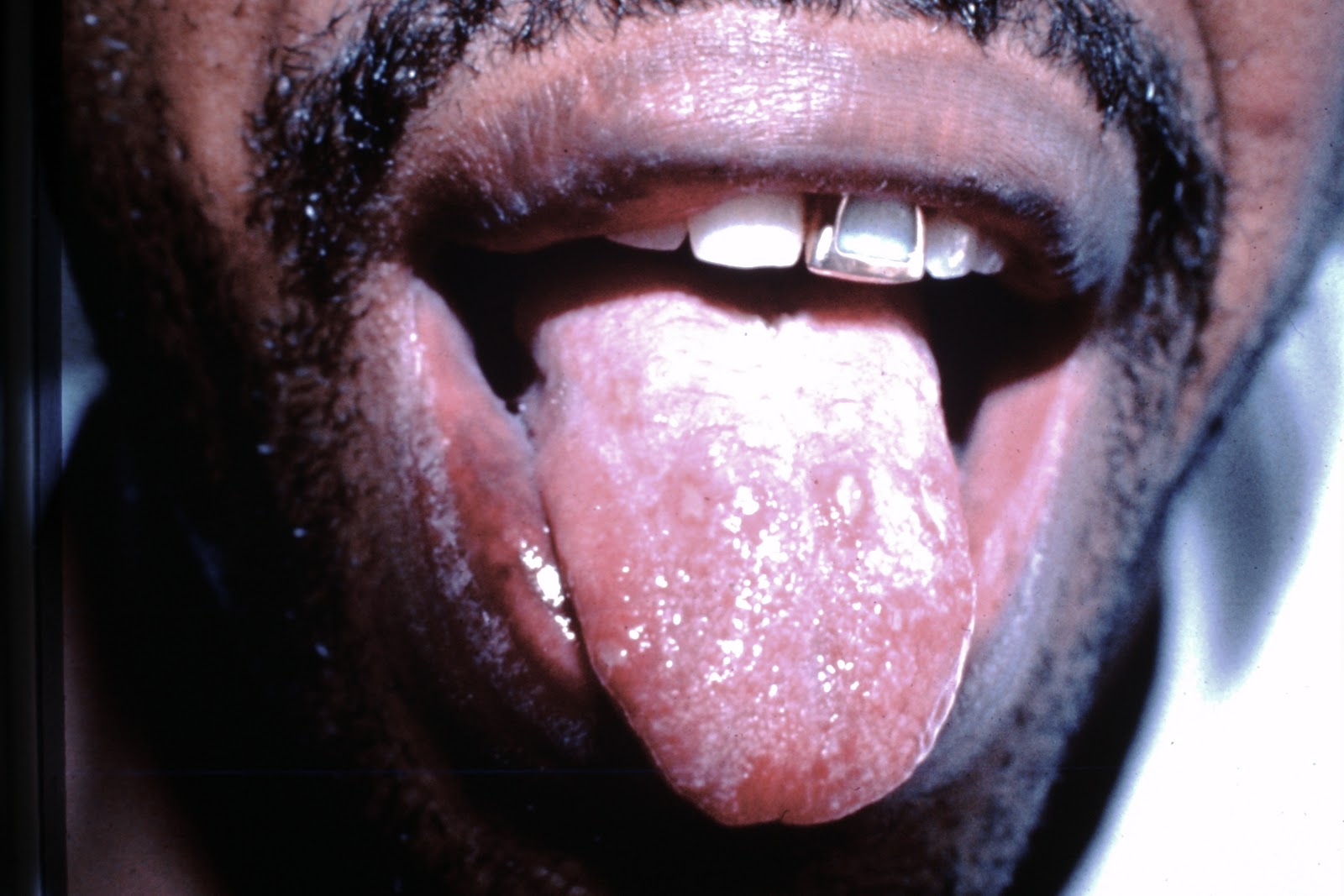
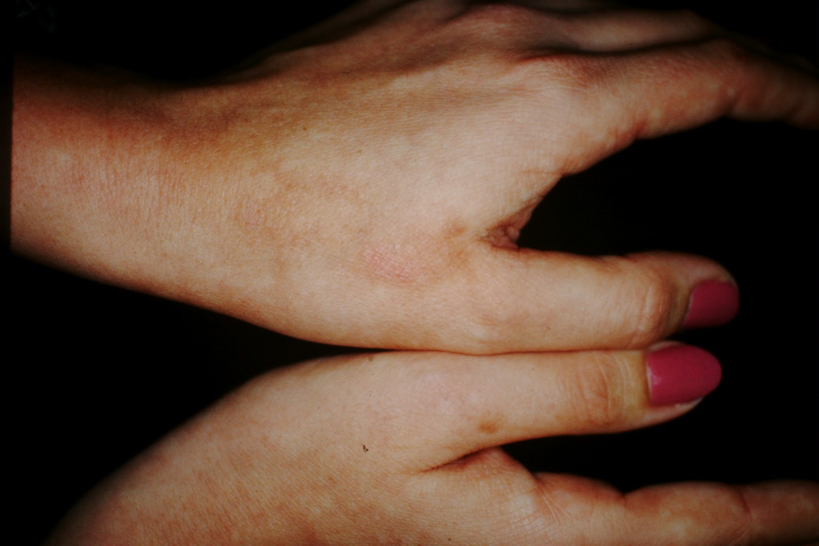
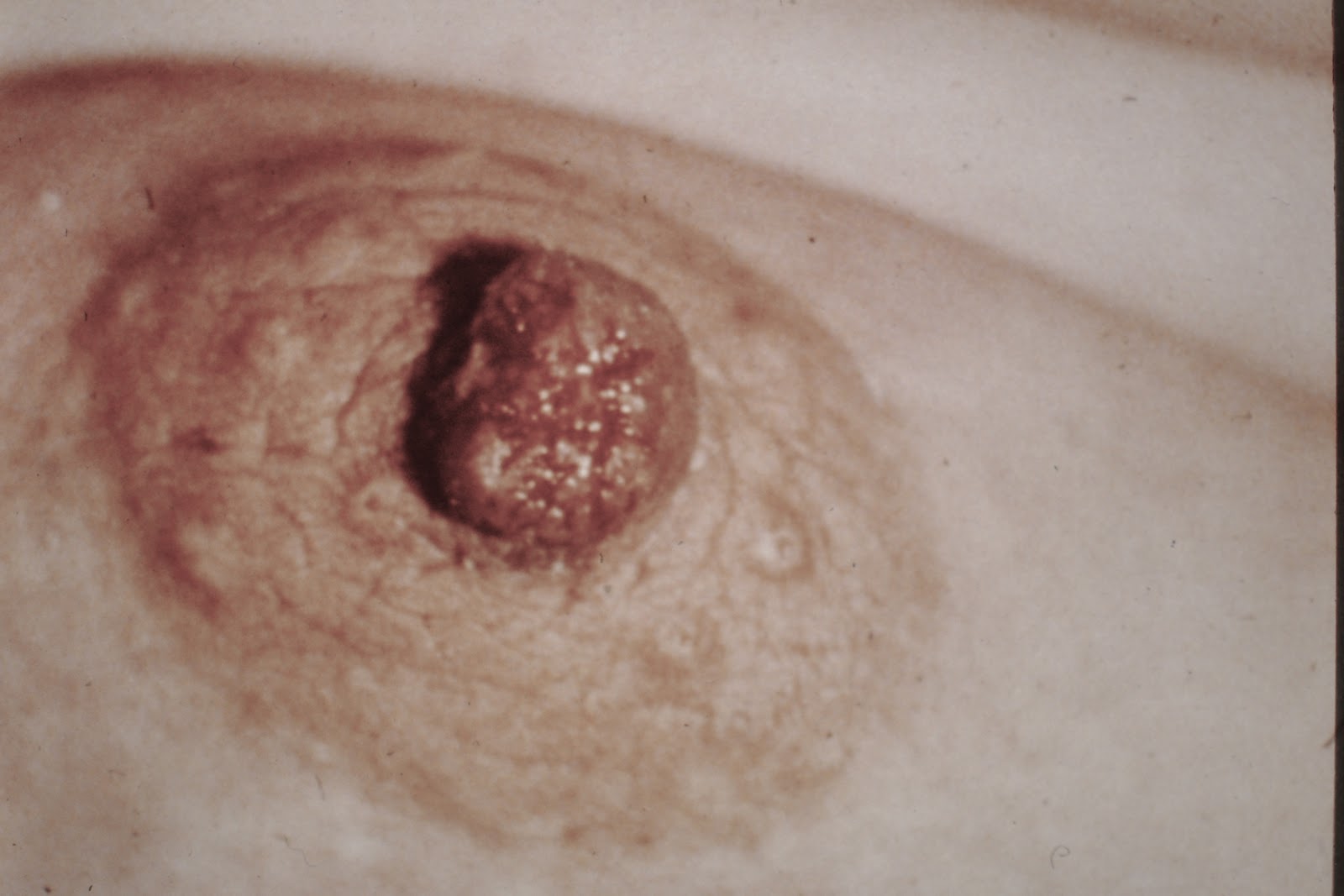
Of course, the last Q was CREST, a variant of scleroderma, and from my description there is no way you could tell whether or not it was systemic scleroderma or merely CREST, so you got credit for either answer.

Several of you wanted to know a little about me, so I will fill you in on who I am and why I chose to do the Challenge.

As most of you know, while I was in residency, I managed to amass a collection of clinical images which rivaled many of the academic programs with which I had been associated, and I always felt I needed to share them with my PAs, in a somewhat controlled way which would enhance learning and engender a desire to learn more.  After casting around a bit, I hit on the idea of using wine as a reward, because it coincided with my love of wines and gastronomy in general.  

After a while, we generalized it to other PAs and ARNPs, which eventually ended up with our blog getting in the neighborhood of 25,000 hits from all parts of the US and the world.  Even Romania and Bulgaria!  It turned out to be a lot of fun from my perspective, because I got to know some really bright, motivated people along the way, and it also honed my knowledge base a bit.  I've always enjoyed general as well a surgical derm, although 90% of my practice is related to derm onc, and it forced me to remember a lot of details which otherwise would soon slip into the part of my brain devoted to baseball statistics and differential equations.

I am happily married and have a daughter Liana (seated on the couch above); her boyfriend Josh (in plaid); a son Daniel (blue shirt, who has been a subject of a couple of Q's); a daughter in law Caroline who is Quebecoise; and a marathon-running, five bar exam passing, bicycling, book writing wife with whom I spend on average an hour a night, drinking wine and talking about the world.  What a family.  What a life...

My hobbies are modernist cuisine, wine and bicycling, and I am currently preparing to ride all of the significant climbs of the Tour de France this July, in the Pyrenees, the Alps and Provence.  I am training with Kevin Livingston, who used to be He-Who-Shall-Not-Be-Named's lieutenant on the Postal Team and Kevin now works in Austin at Mellow Johnny's, and I am lucky to have him as my coach.  He's a bit of a sadist, though.  He says it's for my own good.  We'll see.  

And, for the record, I truly believe what Lance did was no better, but no worse than any other cyclist of his era.  He was just better at it, in all phases of both riding and doping, and so he became the whipping boy.  And his personality is a bit strong, to put it mildly, so that also had something to do with it.  It is well known that the most widely admired cyclist of all time, Eddy Merckx, used to take amphetamines prior to the mountain stages, which was a hell of a lot more dangerous than EPO and Testosterone.  

So, for those of you who struggled and swore and pored over your texts (and Google), I have both admiration and a sense of kinship.  You struggled, and the knowledge you walk away with is not only the specific (e.g. how to diagnose PCT) but also more important, you learned that between what you know and what you can access, there is virtually nothing you can't figure out.  Your patients are very, very lucky to have you.  

Lastly, BE YOUR OWN BRAND!  If you do, you will always, ALWAYS have a way to make a good living, and you will be immeasurably important to those for whom you care.

Dr. B

Pyoderma Gangrenosum

PG is, to say the least, an exasperating disease, both from the standpoint of the patient as well as the treating provider.  It can be refractory to every treatment in Shelley and Shelley, or it can heal on its own, or it can respond one time to one regimen, then be refractory the next go-round.  

PG is notorious for, in its smaller iterations, being confused with spider bites.  If you EVER hear of anyone who says they get multiple spider bites with subsequent necrosis, nine times out of ten, you are dealing with PG.  

The hallmarks of PG are all there for you in the pic above:  Purplish, undermined skin, evidence of precious ulceration, and the history of pathergy, that is a pathologic response to trauma.  Think of it like the Koebner Phenomenon of psoriasis; it is an abnormal physiologic response to external trauma.  

Necrobiosis Lipoidica is a different animal in that it tends to have much smoother plaques that are more uniform, and tend to be more pretibial, although that is not always the case.  

If you answered correctly, then click on this link and enjoy my favorite song from the eighties : http://www.youtube.com/watch?v=EHJmPcILfg8

If, however, you didn't, punish yourself by listening to this link, which is possibly the worst song ever recorded : http://www.youtube.com/watch?v=-rfD8NQLO8w


Wednesday, December 12, 2012


Late Phase Porphyria Cutanea Tarda

This was a toughie for a lot of you, and so I will describe what I see which, in my mind, are the critical cues for making the correct diagnosis.  What I see are multiple erosive-looking scars on the dorsum of the hand, with a milium on the fourth digit.  Any time I see any changes, either blisters (which I rarely see) or exaggerated aging, which I commonly see, or scarring, as above, I think the patient has PCT or pseudoporphyria until proven otherwise.  

As you all know, PCT has a predilection for the dorsal hands and, along with other findings of PCT (name three for three bonus points) is a marker for internal disease.  Usually, it is associated with an underlying condition, with hemochromatosis and alcoholic cirrhosis being common factors, but by way of pathogens, Hep C and HIV are the usual suspects.

Why wasn't this LP?  Well, I have seen LP just about everywhere, but the dorsal hands are not a really common location.  I like the wrists, legs, lower back, nuchal scalp, etc., but dorsal hands, not so much.  So, once again, if you see changes on the dorsal hands, think PCT.

Monday, December 10, 2012


Linear Scleroderma en Coup de Sabre

Linear scleroderma is a condition characterized by localized linear atrophy that resembles the findings one would find, at the end of the Franco-Prussian War, in soldiers who had been injured from cavalry charges from the saber-wielding Prussian Cavalry.  Since this description is in French and not in German, one would correctly presume the French bore the brunt of these kinds of injuries.

The onset of this condition is, as above, in childhood and usually is absent of underlying neurological deficits, unlike its cousin Parry Romberg Syndrome, which is a more severe condition involving progressive facial hemiatrophy, which can result in the entire affected side of the face being smaller than the other.  On a clinical basis, without this difference from side to side, one would have to lean on en Coup de Sabre being the preferred diagnosis.  This is just such a case.  

Linear Scleroderma en Coup de Sabre is usually unaccompanied by routine laboratory abnormalities, except in research settings.  Thus, although Scl-70 and ANA titers can be out of whack, usually they are not.  In fact, some authors question whether this entity is at all related to morphea or scleroderma.  

This disease does not, in its pure form, extend below the orbital ridge, although there are overlapping cases with Parry Romberg which go lower.  If you study the case above, you can see the atrophy, the superficial nature of the vasculature and the linear nature of the defect.

Sunday, December 9, 2012


Fabry's Disease

Trust your eyes.  When you see something that looks like cherry hemangiomas, trust your eyes.  Many of you ended up working this question backward, by trying to put something together that had both skin lesions and urine pathology, and came up with Henoch Schoenlein Purpura, which, of course, looks nothing like these angioma-like structures in the top picture.  You didn't trust your eyes.  

Fabry's Disease is an X-linked disorder which leads to deposition in the endothelium of a certain kind of lipid which leads to the pathophysiology.  Many times the disease manifests itself with symptoms of a CVA, or renal insufficiency, or myocardial infaction in a relatively young person, and if it is in association with these lesions we see above, known as angiokeratomas (which look  a  lot like cherry angiomas) then one should consider Fabry's Disease.  The finding is classically called angiokeratoma corporis diffusum.  

The classic finding in the urine is the lipid droplets, as seen above.  It is rare that derm will be the first to make the diagnosis of Fabry's but at the same time we often times can help the other specialties tie it all together.


Thursday, December 6, 2012


Primary Syphilis

Given that the answer is "no" to the question you should be asking (which is "does it hurt?") the differential diagnosis is mercifully brief.  A large, painless ulcer on the penis?  There is one and only one answer.  Our little treponemal friends are just setting up shop, preparing for what they hope is a long, fruitful (from their perspective) relationship with Mr. Happy's friends including the skin, the brain, the spinal cord and eventually the deep tissue.  

Wednesday, December 5, 2012


Disseminated Intravascular Coagulation


The way to think of this problem is not so much to look at it and say "DIC" but rather to look at it and say to yourself "This is clearly a vascular problem which resulted in small vessel obstruction, followed by bleeding into that same compromised vascular distribution".  As such, you would come up with a ddx which would, eventually, include DIC.  

Think of DIC as not really a disease, but rather a manifestation of disease, whether it be from sepsis, trauma, systemic disease (e.g. pancreatitis), malignancy associated thrombotic changes, envenomations from certain snakes, or anything else which could trigger the coagulation cascade (abruptio placenta, weird manifestations of antiphospholipid syndrome, etc.)

On a localized level, therefore, I would include frostbite, which most of you listed, as one of these phenomenae, so don't feel bad for listing it.  I've seen frostbite that looks just like this, and although I have never seen DIC in person, I suspect there would be plenty of other clues to figure that out.  I also doubt I would have the presence of mind to take a pic of DIC unless, like above, the patient survived.

A few of you listed DIC and one of you (you know who you are) listed ONLY DIC, which was a pretty bold move.  

Interstingly, the inflammatory cytokines seen in certain trauma patients mimic the profile seen in sepsis, and the current theory is that it is the cytokines which trigger the cascade leading to DIC.


Tuesday, December 4, 2012


Cholesterol embolism

Cholesterol embolism usually occurs after invasive vascular procedure, and it marked by discoloration of distal circulatory fields such as the pads of the toes, as above.  The blotchy, discolored appearance, especially if sudden in onset, is referred to as "blue toe syndrome" and is very highly suggestive of cholesterol emboli.  Some of you noted the pincer nails and onychomycosis, but the reality is, given the clues all of you should have honed in on the vascular hints and nailed this one.  Pun intended.  

Monday, December 3, 2012


Pruritic Urticarial Papules and Plaques of Pregnancy

Pruritic Urticarial Papules and Plaques of Pregnancy, aka PUPPP, is a benign condition that arises classically in the third trimester for a primiparous female.  Of note, a common dermatosis of pregnant females is atopic dermatitis of pregnancy, which occurs earlier in the pregnancy.

The classic description of PUPPP is the development of intensely pruritic urticarial papules within the striae of a primip.  HOwever, in some cases, it can occur in multips, and the lesions may not be confined to the striae.  Typically, there are no bullae, but there may be very small vesicles.

Of course, the IF is negative in PUPPP.  It would be positive in pemphigoides gestationis, but is never positive in PUPPP.


Thursday, November 29, 2012


Urticaria

I'll keep this short and sweet:  If you want to distinguish between urticaria and urticarial vasculitis, either ask the patient if the lesions last more than 24 hours, or have them come in after having drawn a border around the urticarial lesion, much the same as if you were documenting cellulitic change.  As one of our playas noted, you can also look for petichiae or bruise-like change to sniff out UV, but the easiest way is simply to ask the patient how long they last.

Wednesday, November 28, 2012


Rocky Mountain Spotted Fever

Rocky Mountain Spotted Fever is a rickettsial disease caused by Rickettsia rickettsii, and the natural history of RMSF goes like this:  1-2 weeks after being bitten by a tick from the Ixodid ticks, primary among them are the wood ticks (Dermacentor andersoni) and dog tick (D. variabilis), the victim develops constitutional symptoms (fever, chills, malaise) and a centripetal rash that begins on the distal extremities and marches inward toward the trunk.  This time lapse from the onset of the rash to the truncal manifestations can be as short as 6 hours.  Over the next couple of days, the rash becomes petichial and hemorrhagic, and if the patient isn't treated by day 5, the fatality rate rapidly climbs.  Problem is, in up to 20% of cases, there is no rash.  The usual cause of death is acute renal failure. If treated in time with a tetracycline derivative or chloramphenicol, the death rate is nil. 

About four years ago, I  had the pleasure of having a different rickettsial disease, R. conorii being the causative organism (bonus: which disease did I have and where did I get it?) and I thought someone was jackhammering the inside of my skull for about a week, in spite of starting Doxy as soon as I figured it out.  Not fun.  

Tuesday, November 27, 2012


Lupus pernio (Sarcoidosis)

First off, this is a testmanship question.  I just discussed unknown rashes, etc. on African American patients being sarcoidosis until proven otherwise, and then I throw out an image of strange papules on the nose, and- imagine that!- he's African American.  You could win lots o' dough in Vegas if you were to hit the tables with these kinds of odds in your favor.  

Lupus pernio is characteristically violaceous to skin colored, smooth shiny papules and plaques around the nose, cheeks and lips.  It is associated in nearly 75% of cases with chronic fibrotic respiratory tract involvement, so the correct diagnosis is important to the patients because the correct treatment can interrupt a cycle of disease that may cripple or rarely kill the patient.  Lupus pernio is associated with other manifestations of sarcoidosis, including bony involvement, but the primary risk is the association of the pulmonary tree.  

For those of you who got it right, reward yourself by clicking this link:  http://www.youtube.com/watch?v=9bZkp7q19f0

If you missed it, click this link:  http://www.youtube.com/watch?v=pWS8Mg-JWSg

Monday, November 26, 2012


Sarcoidosis

This is a pattern-recognition question.  If you take away the patterned aspect of this Q, you end up with a laundry list of ddxes: everything from infectious (deep fungal, AFB, bacterial, parasitic, viral) to neoplastic (weird CTCLs, etc.) to factitial (excoriated bug bites, etc) to inflammatory.  This is, of course, a special kind of inflammatory condition, and one that is all too common in African Americans.

From now on, for the rest of your natural lives, please remember this:  Any weird rash on African Americans can be sarcoidosis, and the importance of this point is this:  In many cases the skin manifestations of sarcoidosis precedes the other symptoms, and as such is the bellwether for the disease.  The worse the skin disease, the worse the systemic sarcoidosis.  And since we are often tempted to treat inflammatory skin diseases empirically, we can miss an important cutaneous manifestation of a systemic disease.  Given that sarcoidosis can irreversibly damage kidneys, eyes, lungs and liver, it is well worth recognizing the possibility and diagnosing it before the cat, so to speak, gets out of the bag.

Sarcoidosis, like syphilis, is a great imitator.  It can look like many different diseases (see the first paragraph above) and can cause great harm, like lues, if left unrecognized. 

Sarcoidosis is more common in the African American community at large (three times as common as the general population) and is more severe than in Caucasians.  So, in the future, if I throw up an image, and it is from an African American, and you can't tell exactly what it is, your answer should be "sarcoidosis until proven otherwise".

Sunday, November 25, 2012


Allergic Contact Dermatitis to Gear Shift Knob (Australia)

This was, no doubt, a challenging question.  The problem with trying to piece together a story for a guy with a localized contact derm on the hand is that you need some history to pull everything together.  If you practice is like ours, most of the time the patient does not present to your office with a known allergen in mind, because if they did, they wouldn't be bothering you with a mystery they've already solved.

In this case, I wanted you to put it all together.  Your clues were what you see above, which is a clear case of localized allergic contact dermatitis.  Second, I gave you some clues:  1) He was a skin cancer patient  2) It was a localized dermatitis secondary to rubber.  At this point (as one of you did) you should be thinking "why did he say skin cancer patient?" and put together that it had to be in a latitude conducive to skin cancer.  But wait!  There's more!  Why the localization to the left hand?  Gotta be an answer.  That was when our Sherlock figured out that in some countries that were formerly part of the British Empire (or influenced by them as in the Japanese) they drive with the shifter on the left, not on the right.  The British Isles are not known for their skin cancer problems, so it had to be either Australia,  or New Zealand.  Playing the odds, since Australia is much bigger than New Zealand, the answer was Australia.  

Australia has a very highly developed derm community, and first among the superstars of Aussie derm is Robin Marks. For a bonus point, what does "Slip, Slap, Slop" mean? Dr. Marks has developed a body of literature which has informed the rest of us on such subjects as which side of the body has more AKs and what is the likelihood an individual AK will turn malignant.  Not bad for a country founded as a penal colony...

Monday, November 19, 2012


Behcet's Disease

Behcet's is a pretty interesting syndrome, in that it affects widely separated parts of the body, and tends to be pretty serious.  Also known as Oculo-Oral-Genital Syndrome, it consists of recurrent aphthous oral ulcers at least three times in twelve months, in conjunction withany two of the following:  retial vasculitis or uveitis, cutaneous lesions such as erythema nodosum, pseudofolliculitis or papulopustular lesions (think lesions similar to fire ant bites), a positive result of a pathergy test or recurrent genital ulceration.  A positive pathergy test is the stimulation of the formation of similar lesions by scratching or puncturing the skin.

The proper way to pronounce Behcet's is Beh-Chets, NOT Beh-shet's.  I would have allowed Beh-Jet's, since the ch sound in Turkish is similar to a j, but nobody answered that way.  


Thursday, November 8, 2012


Tuberous Tendinous Xanthomas

Well, this is a repeat question (from May 16th) and so I will direct you to that discussion, but the most important aspect of this question is that many of you got it right.  Please think in terms of depositions into the skin, and then run your differential dx based upon location, appearance and peripheral clues.  You all nailed the elevated lipids, so nice job overall.  By the way, this was repeated in error, and I apologize for not making it a harder Q.  

Wednesday, November 7, 2012


Necrobiosis Lipoidica

Necrobiosis Lipoidica is a degenerative disorder of collagen, which manifests itself with plaques that are typically erythematous to yellowish in the center, fading to erythematous to violaceous at the periphery.  The usual age on onset is in the thirties, it usually (but not always) accompanies glucose metabolism dysfunction, and it is more commonly than not on women.  The most common laboratory abnormality is an abnormal HgbA1C, but often they also exhibit an elevated fasting blood glucose. 
The lesions can ulcerate as a result of trauma, and can be precipitated by trauma.  The clues to diagnosis are location (pretibial), color (that characteristic yellowish color in the center of the lesion gives it away) and the pertinent negatives: it is not ulcerated, it is not focal like an infection and it is not elevated, as in pretibial myxedema.  It is not annular, like GA, either. And, unlike Erythema Nodosum, it is multicolored and shiny in the center, as opposed to EN which is more focal, usually uniformly red and, if anything, slightly raised. This is a bit atrophic.
As mentioned above, it is more common in diabetics, and used to be known as Necrobiosis Lipoidica Diabeticorum, but  now, since it is not just associated with diabetes, it is simply known as Necrobiosis Lipoidica.

Tuesday, November 6, 2012


Granuloma Faciale

Granuloma Faciale is a benign inflammatory process which results in elevated papules, nodules and plaques, nearly always on the face but occasionally on other sun-damaged skin.  The lesions can be skin-colored, brown, or erythematous to violaceous, are smooth with a "peau d'orange" appearance.  

Pathology (obtained with a punch biopsy) shows a characteristic Grenz (pronounce "grents") Zone which translates to a clear area between the inflammatory infiltrate and the skin.  It is almost as if the infiltrate were epidermophobic.  One can also see a Grenz Zone in Leukemia Cutis, Cutaneous B-Cell Lymphoma and Lepromatous Leprosy.  


Sunday, November 4, 2012


Sous Vide Cooking

Sous vide (pronounced "sooveed") literally means "under pressure", but in reality refers to cooking foods to their optimum temperature to allow the optimum flavor to develop, texture to be displayed and for nutrition to be maximized with any given dish.  First developed in the 1970s by the food scientists who work for the French National Railroad (SNCF), it languished in long-forgotten cookbooks until the late eighties, when a culinary genius named Ferran Adria began using sous vide cooking in his fantastical preparations at his restaurant outside Barcelona named El Bulli, for his two favorite pit bulls.  

Other chefs quickly followed, most notably Thomas Keller from the French Laundry and Per Se, Heston Blumenthal from The Fat Duck, and Grant Achatz, more recently, from Alinea in Chicago.  It is now considered to be a standard form of cooking in nearly all of the best restaurants in the world, for the reasons listed above.  

The former Chief Technology Officer at Microsoft, Nathan Myrhvold, growing bored with his corporate life, bailed out and went to work for Heston Blumenthal, who currently holds the crown as the best restaurateur on the planet, believe it or not as a prep boy, then sous chef.  Anyway, he gleaned enough information from that experience and collaborating with others, including Adria and Keller, to write a fifty pound, five volume  cookbook called Modernist Cuisine.*  

Well, at the same time all this intellectual ferment was taking place, I was in a funk from a cooking standpoint, because many of the great meals I had recently had were not reproducible in my home.  Daniel Boulud, Keller, Charlie Palmer and others were, from a technique standpoint, killing me.  So, when Myrhvold came out with MC, I jumped on it, reading it cover to cover figuring out what I had been missing. Mostly, as it turns out, it was techniques that were dependent upon equipment I didn't have, and chief among those techniques was sous vide cooking.

After researching my choices, I bought a Sous Vide Supreme water oven, and became an instant convert.  The first four meals I cooked were the best chicken, duck, shrimp and fish I had ever eaten in my life, and it was at home, not in a 5 star restaurant!  Now, my wife Cindy and I cook 90% of our main courses using sous vide techniques.**

Here's what you all really want to know:  The reason sous vide cooking works so well is that at roughly 154 degrees Fahrenheit, collagen starts to foreshorten, which causes the tissue you are cooking to contract, which then expels both flavorful ingredients as well as liquids- the juices you try so hard to preserve in your meals. If you cook the meat instead at 140 or even lower, for a longer period of time, you will find the flavors remain intact, the juices remain in the meat and you end up with a far superior culinary result.  

Equally amazing is that if one cooks, say, a tough cut of meat such as a brisket or short ribs, for a long period of time (48 to 72 hours) the collagen becomes gelatinized which adds both to the flavor and the texture of the food.  

In the last half decade or so, we have been able to be our own publisher and PR agent (via Facebook), we can distribute original video productions (YouTube) and now, we can literally reproduce world class cuisine at home, with a modicum of new equipment.  Now, that's cool!

*Myrhvold now has a more accessible version of MC called Modernist Cuisine at Home.  It is markedly less expensive, more accessible and the recipes use equipment that is more reasonable than the high priced gadgets featured in MC.  

**I now have three different options for cooking sous vide, and at times use all three of them to prepare multicourse meals.  


Wednesday, October 31, 2012


Frontalis-Associated Lipoma

In 1989, Stu Salasche, Bill Grabski, Martha McCollough and Vince Angeloni described an entity they described as being either enveloped in the frontalis muscle of the forehead, or just beneath it.  Their term, Frontalis-Associated Lipoma, has stuck with this entity ever since.  

The tricky thing about FAL is they are not easily removed, and in fact are a real pain in the neck because of the investiture in the frontalis.  My preferred technique, which involves a horizontal incision following the rhytids, followed by significant undermining and then a vertical incision in the frontalis, following the muscle fibers, down to the lipoma, is by far the best way to handle them.  I learned that technique from the authors themselves, while they were on faculty at our sister residency at Brooke Army Medical Center in San Antonio.  

The clues that we were not dealing with an epidermal inclusion cyst are primarily that the lipomas are soft and much more mobile than a cyst of that size.


Tuesday, October 30, 2012


Leukemia Cutis Secondary to CLL

This very nice gentleman is a patient of mine who has been with me for well over a decade, and he presented about two weeks ago with a nodule on the scalp which was biopsied and found to be a squamous cell carcinoma.  Interestingly, though, my pathologist (she is very good!) noted sheets of lymphocytes around the lesion and asked me if, perchance, our patient had CLL.  The answer, of course, was yes, but it was well controlled.  Up til now, that is.

We scheduled him for surgery within two weeks, as we do all invasive SCCs, and when he came to the office for the surgery, he mentioned he had some swelling on his forehead for the last couple of days.  The plaques were slightly erythematous, firm to the touch and warm.  Well, the differential diagnosis in my head really came down to infectious versus neoplastic, but his CBC showed 20K lymphs with zero activity going in the PMN range, but a vast predominance of lymphs. This makes the diagnosis of leukemia cutis a virtual certainty.   Had it been infectious, we would have seen a much less lymphocytic, much more PMN-rich differential. Additionally, we had the comments from the pathologist regarding the SCC to base our opinion on.

Leukemias are proliferations of clonal populations of leukocytes (in this case lymphocytes) which are maturationally arrested. Leukemia cutis represents clonal proliferations in the skin which may be seen in AML, ALL, CML, CLL, as well as hairy cell leukemia and myelodysplastic syndrome.  If it is associated with AML or ALL, it has a very grim prognosis.  


Monday, October 29, 2012


Palmoplantar Keratoderma

Palmoplantar keratoderma, aka Keratosis Palmaris et Plantaris, is a not really as much a diagnosis as it is a symptom of another underlying condition.  It is, as you see, characterized by a thickening of the palms and soles which may be diffuse or, as above, not.  It can also be punctate, but in my mind that condition doesn't really fit with the others.  

It may be associated with underlying malignancy, resulting sometimes in bizarre patterns such as tripe palms; it may be associated with ectodermal dysplasias, or the straightforward genetic maladies which are too numerous to count in this forum.

In an acquired setting, it can be associated with menopause, meds, infections and systemic disease.


Sunday, October 28, 2012


Pinot Noir

Pinot noir is a finicky, thin-skinned grape which only grows in a fairly narrow climate, which means that it is the high-maintenance cousin of happier, more well adjusted grapes such as Cabernet and Shiraz.  This essentially means that there are inevitably going to be good years and bad years for pinot noir, and so it pays to know whether the wine you buy is from one of those good vintages.

Pinot is grown in a major way in two areas of the US, Chile, Argentina, South africa, Australia and the Burgundy region of France.  For a bonus point, where are the best Cabernets from in France? The only serious Pinots are from Cali, Oregon and Burgundy.  All the others are pretenders.

As you can tell from this pic, taken at Domaine Drouhin winery in Oregon (Domaine Drouhin 1991 pinot was the first Oregon wine I ever drank, on August 14, 1994.  Don't ask me how I remember this.  I am the Rain Man of fine dining.  I remember everything.)

My favorite wines from Oregon come from Domaine Serene, especially their Evanstad Reserve, but overall my favorite of the winemakers is Laura Penner-Ash, who makes both an outstanding Pinot and also a brilliant Shiraz.  

If you ever go to the Willamette Valley, I highly recommend two things:  The Allison Inn is one of the most striking hotels I have ever stayed in, and the Joel Palmer House, a resto that specializes in wild mushrooms.  One of my foodie friends and I had their mushroom tasting menu, which was insanely out of this world.  

Well, enough about epicurean delights.  Time to close out October.  Three more days to weed out the wheat from the chaff, the pips from the grapes.

Thursday, October 25, 2012

Alkaptonuria

Alkaptonuria is an autosomal recessive disease that is a result of abnormal phenylalanine and tyrosine metabolism, characterized by excretion of large amounts of homogentisic acid (aka alkapton) in the urine, hence alkaptonuria.  The circulating HGA causes significant damage to all classes of cartilage (ergo damage to joints and heart valves), leads to kidney stones and a characteristic calcification of the annulus of the intervertebral disks which leads to a syndrome similar to bamboo spine.

It generally is either noted in infancy with discoloration of the urine, or, if undetected then, in the late thirties or early forties, when accumulation of the pigment begins to stain the sclera or, in advanced cases, the tendons.  It also can declare itself, as in this case, by chromhidrosis.  It can be excreted in the sweat glands of the face, the axillae or the areola.  

Now, to the nitty gritty.  I gave you all clues that the meds were not the problem.  I also mentioned she had a stone cold normal examination, including her axillae which were normal as pumpkin pie.  That implied her sclerae were normal.  All this means we are catching her on the front end of the clinical expression of her disease.  For two bonus points, which two countries are noted for high incidences of alkaptonuria. Finally, and most critically, I mentioned (in a separate email, which should have told you it was very, very important) she was in her late thirties.

Which question should you ask her?  Most importantly, you should ask her about dark urine, which she denies.  My next question, which was answered with a "how the hell did you know that?" look was, did she have back problems.  In fact, she had just gone to a back doc the week before and had her MRI the day before I saw her.  She was sitting rigidly straight, as if she were HLA B-27 incarnate.

Labs?  Urine HGA and a CBC, to chase down the rare bleeding diathesis.  If, for some reason, she were negative on the HGA, I would then start looking at heavy metal assays (trying to link the chromhidrosis with the tremor).  


[edit]

Wednesday, October 24, 2012


Delusions of Parasitosis

The Ant
by Ogden Nash

The ant has made himself illustrious
Through constant industry industrious.
So what?
Would you be calm and placid
If you were full of formic acid?


If you really want to cast a curse on someone, instead of wishing that they might live in interesting times, as the Chinese do, you may wish they would develop delusions of parasitosis.  It's a nightmare for these patients, because they so completely believe that the lint, or hair follicles they pull out, or the occasional unlucky stray ant are imbedded in their skin, and they obsess  about them,  which drives them batty. Or, to be more accurate, battier. And they can't understand why you think they're wrong.

Examination reveals linear excoriations, but no primary lesions.  Often, they go from amateur to pro and develop prurigo nodules and ulcers.  This can be the only symptom of psychiatric disease, or it can present secondarily to bipolar illness, schizophrenia, anxiety disorders, and most commonly with obsessive states such as OCD.  

It usually occurs in white middle-aged or older women, although the condition has been reported in all age groups and in men.  Morgellon Disease describes a condition characterized by the delusion of fibers being attached to the skin. Morgellon's has become a new designation for delusions of parasitosis.

Tuesday, October 23, 2012


Median Canaliform Dystrophy

MCD is a condition wherein the nail matrix (NOT nail bed, NOT nail plate, NOT proximal nail fold) is damaged, Often through trauma, but it can also be idiopathic or can be related to genetics.  Think of the nail as a rainbow.  It is formed as ROYGBIV and arcs across the sky, but always the orientation remains the same: ROYGBIV.  So, when you have a MCD as above, the defect is at G (in other words, pretty much in the middle of the nail matrix).  This patient also has a paronychia, but that was a red herring.  

The weird thing about these defects is that they often wax and wane, and may disappear, only to recur years later.  


Sunday, October 21, 2012


Merkel Cell Carcinoma

It's kind of humbling to realize that this cancer was first described in 1972 by Toker.  The Merkel Cell is a pluripotential cell that is of neuroendocrine differentiation, and as a malignancy it tends to be rapidly growing, tends to pick elderly patients and often leads to metastasis and death.  

Nearly half the time it is on the head and neck, but can be virtually anywhere.  The prognosis is worse than that of invasive melanoma, and it has a high rate of local recurrence (one of my patients currently has that) and distant metastases occur up to 75% of the time.  

The tough thing about this clinically is that the tumors begin innocuously, and then accelerate so that by the time the patient goes from thinking it is a pimple until the time of metastasis is fairly short.  

How best to differentiate this from BCC?  If you look at the margin of this tumor, it has a "collarette" of normal skin extending onto the tumor.  BCC almost never does.  Also, it has none of the pearliness of a large nodular BCC.  Nor does it have the telangiectatic surface typical of large BCCs. 

How about SCC?  This is a much tougher call.  It certainly does not fit the keratoacanthoma picture in that there is no central crater, but a large SCC could look like this, but usually it would be more eroded, and instead of the collarette it would be more likely to be wall to wall.  This is typical of a Merkel Cell both in patient age, in location and in appearance.


Thursday, October 18, 2012


Malignant melanoma Breslow's 0.6 mm, with regression 

When you look at a red octagonal street sign, you immediately know what it is and what it signifies.  The same should apply any time you see a lesion like this.  Not many things scare me.  I'm not afraid of spiders, snakes, or heights but I am afraid of anything that looks like this.  The thundercloud gray to pink look in the center of what can only be a melanoma means only one thing:  Regression.  

As you know, regression means that the lymph node has already gotten a taste of the melanoma, even though it is not very thick, and the lymph node then mounted an attack on the primary lesion.  Even in the case of a non-palpable node, it is a metastatic problem  from the moment regression is evident.  Will the metastasis kill the patient?  Maybe, maybe not, but nevertheless it is there.

All of you recognized this was a melanoma, but if you did not mention regression, you did not get full credit for this answer.  When you see a lesion like this, check their nodes and think about SLNBx and heme-onc referral.  


Wednesday, October 17, 2012


Probable Lid Margin Melanoma

We all see pigmented lesions on the lid margin as a matter of daily practice, and the challenge is in distinguishing between benign and malignant lesions.  This case is illustrative of the challenges which face us when we are confronted by such a lesion.

The patient presented to me yesterday and stated she had the lesion for six months or so, and that it had grown significantly over the last six months.  She had permanent eye liner put in approximately seven years ago.

Since I don't have the path yet, I will give you what I see and you can take it from there.  I see a bluish-black pigmented papule on the free margin of the lid, which looks like it extends roughly 2 mm above the surrounding skin.  It is a bit sunken in in the center, and on the conjunctival aspect of the lid, there are areas of what appears to be regression.  Extending further toward the lateral aspect of the conjunctiva, we see extension of the pigment, blue-black proximally,  jet black distally.  

When dealing with pigmented lesions of the lid, there is one rule which almost always is valid: if it extends onto the conjunctiva, it is bad.  In this case, really bad.

The only serious ddx for this (besides perhaps a cellular blue nevus) is that of a pigment granuloma, but the extension onto the conj is so unlikely in that scenario as to be eliminated as a serious consideration.

I will inform you when the dx is out, but I am at this time only giving credit for r/o melanoma.


Tuesday, October 16, 2012


Erythema Elevatum Diutinum

EED is a disease that looks neoplastic, but is inflammatory in nature.  Most of the primary lesions of EED are shiny, smooth papules or plaques that have the appearance of rubbed warts.  Unlike rheumatoid nodules they are mostly epidermal and dermal in appearance, as opposed to dermal and subcutaneous.  Primarily they are found over the hands and feet, knees and elbows.  

The classic histopathology of EED is a leukocytoclastic vasculitis. Attendant to the LCV is fibrosis which gives the EED papules their firm presentation.  

Associated diseases include HIV, IgA monoclonal gammopathy and other hematologic abnormalities, celiac disease, Wegener's, as well as chronic, recurrent strep infections.

Monday, October 15, 2012


The Papulonecrotic Differential Diagnosis

1. PLEVA (pictured here)
2. Lymphomatoid papulosis
3. Papulonecrotic tuberculid ( a rare form of cutaneous tuberculosis)
4. Arthopod assault

This is an important picture, in that the papulonecrotic ddx should roll off your tongue just as easily as ROYGBIV.  This is one of the fundamental descriptors in dermatology.  Congrats to those who got it right!

Thursday, October 11, 2012


Staphylococcal impetigo

Impetigo can be caused by either staph or strep (GABHS) but these days, more is caused by staph than strep.  Bullous impetigo is mostly a newborn disease, and this is clearly not a newborn.  If it were staph scalded skin syndrome, it would be caused by two different exotoxins, which are generated by both a chromosomal process as well as a plasmid-generated process.

Since nearly everyone got this, I will not belabor the diagnostic cues.  

Wednesday, October 10, 2012


Graves' Disease

Graves' Disease is a veritable treasure trove for dermatologic signs and symptoms.  So many of the signs the old dermatologists and internal medicine docs used to look for are now obviated by routine thyroid panels, well before the thyroid disease declares itself by hair loss, hyperhidrosis, melanoderma, Plummer's nails or the like.  In other words, the lab rats are stealing our thunder!  

Pretibial myxedema occurs in roughly 4% of cases, presumably due to thyroid stimulating antibodies which also trigger production of glycosaminoglycans pretibially, preradially and retroorbitally.  The female to male ratio is 7:1.  Bonus q:  is Graves' Disease associated with hyper or hypothyroidism?  

Tuesday, October 9, 2012


Post Streptococcal Erythema Nodosum 

Erythema (red) Nodosum (painful) is an inflammation of the subcutaneous fat, and is triggered by a variety of circumstances, but most of them involve immune complexes being deposited in the medium sized vessels of the septae of the panniculus.  

The appearance is of a slightly elevated erythematous plaque often with, as you see above, a "vascular steal" phenomenon.  They are, as the name implies, very tender.  The location as well as the multiplicity and the tenderness all are clues as to its identity.

Most cases occur in young adult females, are usually multiple, usually pretibial and can be associated with other symptomatology associated with immune complexes such as joint pain, fever, headache and malaise.  Ulceration does not occur and they heal without scarring.  

Historically, tuberculosis was the most common associated disease, but currently we see it most commonly in association with  streptococcal disease, particularly pharyngitis.  Of course, it can also be associated with gut pathogens such as Yersinia, Salmonella and Shigella; but also a kazillion other infectious diseases (too numerous to be listed here) and also, of course, Sarcoidosis.  However, for two additional points, name two deep fungal diseases that cause EN. Also, the estrogens in oral contraceptives can trigger this condition, as can other meds such as sulfonamides.  

Monday, October 8, 2012


Eczema Herpeticum

Eczema herpeticum is a condition wherein the patient has a preexisting condition such as atopic dermatitis, and then suffers a herpetic outbreak, which subsequently seeds the inflamed, eroded skin with HSV.  This is such a case.  

This patient originally had a herpetic outbreak on the lips, which then quickly generalized to nearly all his skin.  Luckily, he was immunocompetent, otherwise this would have killed him.  He did very well on systemic (IV) Acyclovir.  

The clues to the diagnosis are history, especially the perioral history with the spread like wildfire, and also the primary lesions, which are vesiculopustular.  

Also known as Kaposi's Varicelliform Eruption, eczema herpeticum is a devastating disease that can be life threatening if not treated promptly.  The take home message is this, though:  In a patient with a chronic inflammatory condition such as atopic dermatitis or Darier's, if they also have a history of heprpes labialis (or genitalis) please ensure they are prophylaxed so as not to end up with the disseminated variety of their condition.

Thursday, October 4, 2012


Incontinentia Pigmenti

IP is an X-linked Dominant genodermatosis noted by abnormalities in multiple organ systems.  It got its name from the whorled, irregular distribution of pigment in Blaschko's lines.  It is almost exclusively represented in females, being considered commonly a fatal mutation in males, with the exception of XXY Klinefelter's Syndrome males.  It is considered one of the ectodermal dysplasias, and thus will have many of the manifestations of ectodermal dysplasias.  Weirdly, they have a defective X-inactivation system, which causes both normal genetic expression and mutated genetic expression to occur at the same time in the same individual.

The lesions begin as vesiculobullous lesions, progress to verrucous linear and whorled plaques, progress to hyperpigmentation and then eventually they fade, often to hypopigmentation and atrophy.  Occasionally, these women may not know the diagnosis, and with the extremely subtle findings of some IP adult patients, the only clue may be multiple spontaneous abortions (presumably of male babies). 


As with many ectodermal dysplasias, they can have associated hair, nail, ocular or dental abnormalities, as well as neurologic abnormalities including seizures and mental retardation.

In contradistinction to the group of epidermal nevus syndromes, the lesions of IP are more swirled, and instead of being raised they tend to be flat or atrophic.


Wednesday, October 3, 2012


Superficial Spreading Melanoma with Signs of Regression

Try to imagine what this lesion would look like clinically.  What you see is scattered nests and individual melanocytes which have a "dusty (finely granular) cytoplasm, enlarged blood vessels and some evidence of regression in that you can see clumps of melanin in the superficial dermis.  There is superficial perivascular inflammation as well.  How does this translate to the naked eye?

The scattered melanocytes mean that the edge of the lesion will be more diffuse and hazy rather than clear cut, because the usual well-defined melanocytic lesion has nests of cells, rather than individual cells of pigment production at the edge.  

The pigment incontinence gives it a very, very dark appearance, often jet black, because of the depth of the pigment.  This pigmentary incontinence comes from the lesion being attacked by the immune system, which in turn causes the pigment in the melanocytes to leak out into the dermis.  

The enlarged blood vessels give it a slightly pinkish look at the periphery, and that, along with the inflammatory cells, makes it look slightly edematous.

I gave credit for any answer which involved melanoma, because it was a very subtle call between SSMM and MIS with regression.  

Thursday, September 27, 2012


"Bartender Burns" (Phytophotodermatitis)

We've already covered phytophotodermatitis and so I will not belabor the point, but the clue, primarily on the left hand, of hyperpigmentation in an irregular pattern with an accentuated border should cause you to prod the patient about exposure to citrus fruit and then sunlight, and the most common agent to cause this is Citrus latifolia, the Persian lime.  

Enough of you answered other citrus species that I gave credit for those answers as well.  

Wednesday, September 26, 2012


Allergic Contact Dermatitis from Eyedrops

This is a clinical conundrum which really demands every single bit of your clinical diagnostic skills.  When we think about periorbital skin changes, we think about atopic dermatitis, allergic contact dermatitis and dermatomyositis.  First, think atopic derm.  usually on the lower lids, usually associated with diffuse, poorly demarcated lichenification on the lower lid (with occasional upper lid involvement) and often with other stigmata of atopic dermatitis (such as Dennie-Morgan folds, allergic shiners and nasal creases).  The heliotrope rash of dermatomyositis is in the distribution of the orbicularis oculi muscle (which, incidentally, may be tender because of the underlying myositis), is swollen and pinkish-violet.  It is almost always symmetrical.

This patient has an asymmetrical rash that looks like she suffered from drippage on her left side, it is lichenified, the rash extends well beyond the orbicularis and is far more prominent on the lower lid than the upper.  There are no other stigmata of atopic dermatitis. Given this clinical picture, the best bet would be allergic contact dermatitis.


Tuesday, September 25, 2012


Erythrasma

Erythrasma is a condition of the moist, intertriginous areas of the body which represents a superficial infection of the epidermis with Corynebacterium minutissimum.  It is distinguished from tinea cruris by the more diffuse border and by a more diffuse involvement of the central part of the plaque, as opposed to the more healed appearance of tinea cruris.  

Erythrasma fluoresces coral pink (or red) on Wood's Lamp inspection due to excess coproporphyrin III, which is a metabolic byproduct of the causative bacteria.  Please remember the fluorescence will not be present if the patient recently showered.  It is also important to note the bacteria do not fluoresce blue or blue-green, because the organisms that do that, Microsporum canis and M. audouinii, are the causative agents of tinea capitis, but not tinea cruris.  For a bonus, what is the most common fungal organism to cause tinea cruris?

The vast majority of you killed this one.  Good job.

Monday, September 24, 2012


Mammary Paget's Disease Secondary to Infiltrating Ductal Carcinoma

Paget's Disease of the breast is named for Sir James Paget, who was one of the great surgeons of his time.  He first reported the disease, which he initially believed to be benign, on the nipples and areola of fifteen women.  He reported the connection with intraductal carcinoma in 1881, and the disease is now considered to be a manifestation of underlying ductal adenocarcinoma that invades the epithelium of the nipple, leading to eczematous changes and erosion.

The prime clue in this case is the history that this is a unilateral condition.  Can jogger's nipple occur unilaterally?  Yes, but you better be darn sure that's al it is.  Close follow up is imperative in these cases, and the best way to get to the answer is to biopsy the lesion.  Short of that, an extremely short follow up after conservative treatment would be your next best option.  Me?  I say biopsy it.  

Extramammary Paget's is outside the purview of this discussion, but perianal and perigenital or axillary rashes that do not resolve deserve a biopsy.  'Nuff said.