Thursday, September 27, 2012


"Bartender Burns" (Phytophotodermatitis)

We've already covered phytophotodermatitis and so I will not belabor the point, but the clue, primarily on the left hand, of hyperpigmentation in an irregular pattern with an accentuated border should cause you to prod the patient about exposure to citrus fruit and then sunlight, and the most common agent to cause this is Citrus latifolia, the Persian lime.  

Enough of you answered other citrus species that I gave credit for those answers as well.  

Wednesday, September 26, 2012


Allergic Contact Dermatitis from Eyedrops

This is a clinical conundrum which really demands every single bit of your clinical diagnostic skills.  When we think about periorbital skin changes, we think about atopic dermatitis, allergic contact dermatitis and dermatomyositis.  First, think atopic derm.  usually on the lower lids, usually associated with diffuse, poorly demarcated lichenification on the lower lid (with occasional upper lid involvement) and often with other stigmata of atopic dermatitis (such as Dennie-Morgan folds, allergic shiners and nasal creases).  The heliotrope rash of dermatomyositis is in the distribution of the orbicularis oculi muscle (which, incidentally, may be tender because of the underlying myositis), is swollen and pinkish-violet.  It is almost always symmetrical.

This patient has an asymmetrical rash that looks like she suffered from drippage on her left side, it is lichenified, the rash extends well beyond the orbicularis and is far more prominent on the lower lid than the upper.  There are no other stigmata of atopic dermatitis. Given this clinical picture, the best bet would be allergic contact dermatitis.


Tuesday, September 25, 2012


Erythrasma

Erythrasma is a condition of the moist, intertriginous areas of the body which represents a superficial infection of the epidermis with Corynebacterium minutissimum.  It is distinguished from tinea cruris by the more diffuse border and by a more diffuse involvement of the central part of the plaque, as opposed to the more healed appearance of tinea cruris.  

Erythrasma fluoresces coral pink (or red) on Wood's Lamp inspection due to excess coproporphyrin III, which is a metabolic byproduct of the causative bacteria.  Please remember the fluorescence will not be present if the patient recently showered.  It is also important to note the bacteria do not fluoresce blue or blue-green, because the organisms that do that, Microsporum canis and M. audouinii, are the causative agents of tinea capitis, but not tinea cruris.  For a bonus, what is the most common fungal organism to cause tinea cruris?

The vast majority of you killed this one.  Good job.

Monday, September 24, 2012


Mammary Paget's Disease Secondary to Infiltrating Ductal Carcinoma

Paget's Disease of the breast is named for Sir James Paget, who was one of the great surgeons of his time.  He first reported the disease, which he initially believed to be benign, on the nipples and areola of fifteen women.  He reported the connection with intraductal carcinoma in 1881, and the disease is now considered to be a manifestation of underlying ductal adenocarcinoma that invades the epithelium of the nipple, leading to eczematous changes and erosion.

The prime clue in this case is the history that this is a unilateral condition.  Can jogger's nipple occur unilaterally?  Yes, but you better be darn sure that's al it is.  Close follow up is imperative in these cases, and the best way to get to the answer is to biopsy the lesion.  Short of that, an extremely short follow up after conservative treatment would be your next best option.  Me?  I say biopsy it.  

Extramammary Paget's is outside the purview of this discussion, but perianal and perigenital or axillary rashes that do not resolve deserve a biopsy.  'Nuff said.


Friday, September 21, 2012


Hyperhidrosis

Hyperhidrosis is a psychologically harrowing condition which affects virtually every aspect of its sufferers' lives.  There have been multiple treatments advanced over the years, from using Botox to aluminum salts to meditation and hypnosis.  All of them work to some degree, but the (relatively) recent advent of endoscopic thoracic sympathectomy has revolutionized the treatment of this condition.  One problem that develops in a small minority of patients is that of reflex hyperhidrosis, in which the patient gets compensatory hyperhidrosis which in some cases is dramatically worse than the original problem.  

One of our playas has used a system called Miradry which is an energy based system which eliminates the eccrine and apocrine glands by a combination of heating and cooling.  She reports no compensatory hyperhidrosis in her patients.  It will be interesting to see how this procedure works in the long run.

Thursday, September 20, 2012

Definitions of Lesions

There are many specific descriptors in dermatology which are used to communicate among the cognoscenti regarding lesions.  Among them are vesicle, tumor, scale, crust exudate, etc.  The most basic of nouns related to lesions are papule, plaque and nodule.

Papules are small, less than 1 cm (less than 5mm in my training) and very discrete.
Plaques are broad, raised lesions greater than 1 cm.
Nodules are generally accepted to be greater than 1 cm but less than 2.5 cm, but can include lesions that are up to 5 cm, although that is approaching the range of tumors.

We will have further definitions in our challenge from time to time, but these are the most important.


Wednesday, September 19, 2012


Lymphomatoid Papulosis


Lymphomatoid papulosis (LyP) is a chronic papulonecrotic or papulonodular skin disease with histologic features suggestive of a malignant lymphoma. The disease is characterized by recurrent crops of pruritic papules at different stages of development that predominantly arise on the trunk and limbs. The papules heal spontaneously over 1-2 months, usually leaving slightly depressed oval scars.
This crazy condition has been variously described as a pseudolymphoma, a variant of lymphoma of the indolent T Cell variety, or as a paraneoplastic condition.  Its character, however, is not so much neoplastic as a benign clinical variant.
The condition, as above, comes as both a papulonecrotic and papulonodular variant, and although I have seen both, in my limited experience the papulonecrotic variant is far more common.  
It is imperative that this is a histopathologic diagnosis, with gene rearrangement studies performed to ensure that this does not represent a clonal condition, which would represent a cutaneous T cell lymphoma.

Tuesday, September 18, 2012


Psoriatic exfoliative erythroderma secondary to prednisone withdrawal

I remember very clearly the orientation for my derm residency.  "There are only two things that will get you fired" intoned the Chief Resident. "Coming back from vacation with a sunburn, or putting a psoriasis patient on prednisone".  

Psoriasis is a very, very tricky disease.  It has many different faces (arthritis, plaque type, pustular, inverse, erythrodermic, etc.) and its underlying mechanisms of disease are a complex interplay among different arms of the immune system.  There are, however, some constants.  Psoriasis tends to wax and wane by season, with plaque type psoriasis improving (usually) in the summertime, while inverse psoriasis seems to do the opposite.  

The one thing you can bet on, however, is that if the immune system is systemically suppressed, and if the suppression is not very carefully managed, the patient will respond in an extremely  negative way, with anything from worsening plaques to explosive exfoliative erythroderma.  Since patients who are taking prednisone commonly take themselves off it abruptly or taper according to their own sensibilities, rather than by medical direction, the results can be tragic.  

The clues for today's question were that the patient obviously is suffering from an exfoliative erythroderma, and I told you up front the patient had preexisting plaque type psoriasis.  Many other meds can cause worsening of psoriasis, but if you had to bet your bottom dollar on a case like this, bet it on prednisone. 


Thursday, September 6, 2012


Malignancy-associated Acanthosis Nigricans

Rather than commit a long list of unusual skin findings to memory, I'm going to try to get you to think more in terms of the Zen of the problem.  When you see a particular presentation of, say, acanthosis nigricans, try to put it in a context.  Is it new in onset?  Does the patient have other predisposing issues such as obesity or metabolic syndrome that would explain it?  Is it really dramatic, or less so?  Acanthosis nigricans is a dime-a-dozen condition, but it it is particularly severe, in association with weight loss, or other constitutional symptoms such as night sweats or weakness, you might have a true-blue case of malignancy associated acanthosis nigricans.  Look for adenocarcinomas, in that is the usual combination. 

I am going to list  a few of the signs of internal malignancy, but this list is neither all-inclusive nor will I attempt to describe them.
Leser-Trelat (sudden appearance of itchy SKs)
Bazex Syndrome (acrokeratosis paraneoplastica)
Trousseau's Sign (migratory thrombophlebitis)
Erythema Gyratum Repens
Necrolytic Migratory Erythema
Hypertrichosis lanuginosa
Paraneoplastic pemphigus 
Flushing (associated with carcinoid)
Sweet's Syndrome (Leukemia)
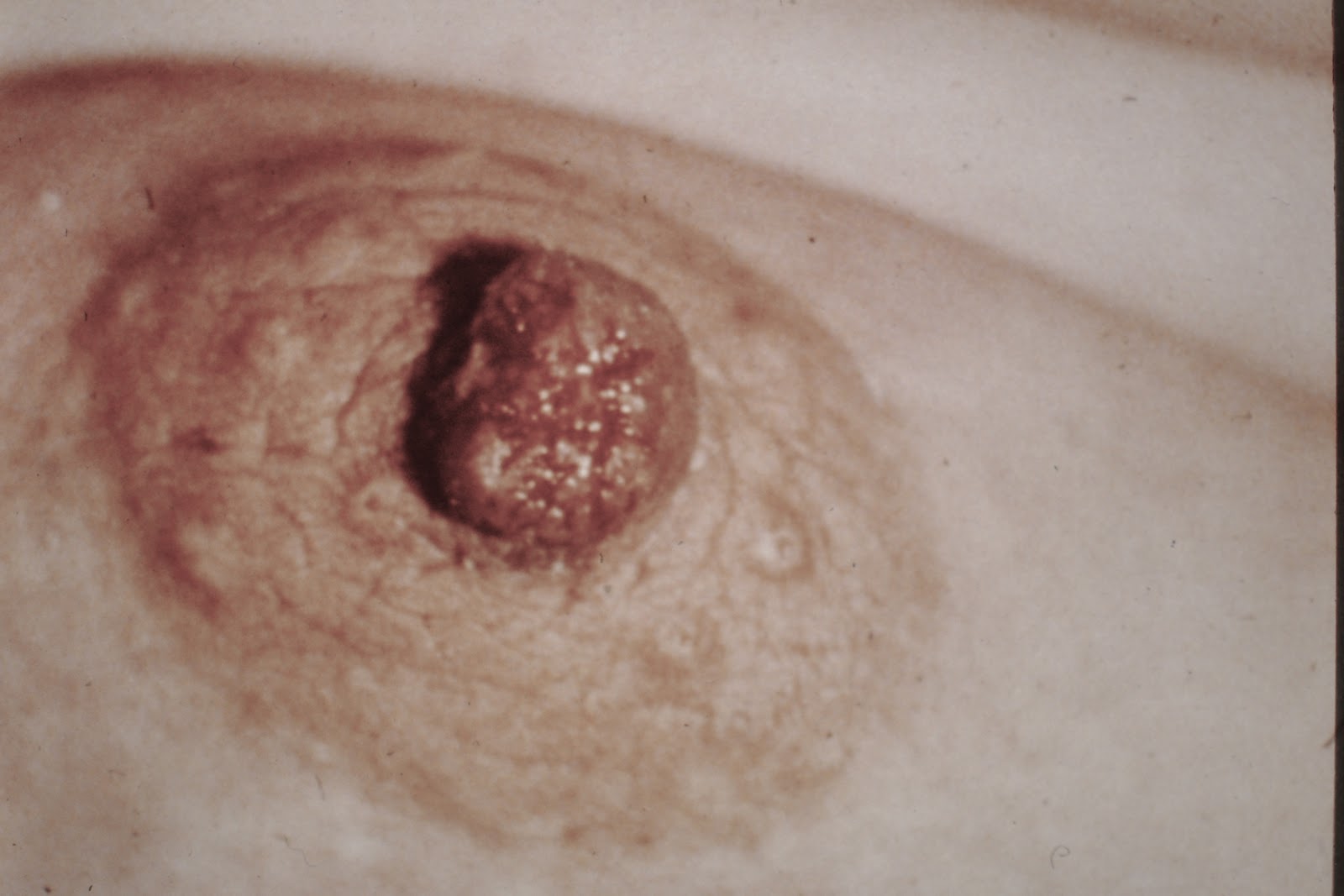
Paget's Disease of the Breast
Extramammary Paget's
Dermatomyositis
Acquired Ichthyosis
Tripe Palms
Sister Mary Joseph's nodule (For a bonus point, who was Sister Mary Joseph?)

Once again, these are not the only ones out there, but if you know these disease presentations, you will often be the smartest one in the room, if not the entire building.  And that's a good thing...


Wednesday, September 5, 2012


Reduviid Bites

“How blessed are some people, whose lives have no fears, no dreads; to whom sleep is a blessing that comes nightly, and brings nothing but sweet dreams.”
Bram Stoker- Dracula


The hemiptera family of insects of which the reduviids are an important group, along with such familiar insects as stink bugs, are an important vector in tropical medicine.  Of the reduviids, the Triatoma are the most important, in that they transmit Trypanosoma cruzi, which is the cause of Chagas' Disease.  These bugs usually hide in the cracks of walls, in the thatching of the roofs and in and under furniture, and only come out at night.  

Reduviid bites are usually unremarkable, with very little in the way of dermatologic manifestations,  but they fall into four broad categories:  papular lesions similar to other insect bites, small vesicles which act as a sort of hypersensitivity reaction, large urticarial lesions with lymphangitis and, finally the erythema multiforme like reactions we see here, which can be either nodular or bullous.  Often these are ascribed to spider bites, but they are not.

The patients rarely remember being bitten, in that the bugs are usually resting on bed linens or the like, rather than on the victim's skin, and use a sucking mouthpart which secretes an anesthetic.  They drink the blood while the patient sleeps, feeding for up to fifteen minutes at a time.  They excrete the trypanosome in their feces, which can be rubbed into the eye, causing a swollen eye called RomaƱa's Sign.  

Tuesday, September 4, 2012


Synblepharon from Cicatricial Pemphigoid

Cicatricial pemphigoid is a chronic scarring and immunobullous disease that is characterized by multiple erosions on the oral mucosa, synechiae on the mucous membranes (including the synblepharon as depicted above) of the eye, mouth, vaginal areas and anus as well as erosions, scarring alopecia and tense bullae on the skin.  

The root cause of CP is autoantibodies which are directed against the basement membrane proteins, specifically the proteins which are found in the lamina lucida.  The usual presentation is one of oral mucosal lesions that have been present for years, with the appearance sometimes of a desquamative gingivitis, and it is most common in elderly females.