Monday, July 16, 2012


Favre-Racouchot Syndrome

Favre-Racouchot is a well-defined syndrome of nodular elastosis with comedones and cysts.  It occurs on the malar and periorbital skin and consists of thickened yellow plaques  studded with comedones and follicular cysts.  It is a result of chronic sun damage, much in the same way that cutis rhomboidalis nuchae, Poikiloderma of Civatte and garden variety solar elastosis all result from too much sun.  Since everyone got this one, I will instead pose a bonus question:  Who (besides me) is the King of the Mountains today?

Sunday, July 15, 2012


Inflammatory Breast Carcinoma with Local Recurrence

One of our great latter day scourges is breast cancer.  In spite of advances in treatment, as well as advances in detection, we still have a relative epidemic of breast cancer, and it still regularly kills women and men.  The picture above is one of tragedy and pain.  This poor woman had a mastectomy some years earlier, and, plague by bouts of depression, ignored the rash that arose around her post-mastectomy scar.  Only after being essentially forced to seek treatment did she get evaluated.  As you can see, the cancer had extended in a pagetoid fashion through the dermis to the surrounding skin and onto the contralateral breast, with an irregular border and with nodules in the field as well.

For those who said radiation dermatitis, please keep in mind that radiation derm is very well demarcated and usually rectangular in shape, with clearly delineated linear borders.  For those that guessed other answers, admittedly this is a tough pic to figure out, but the scar is pretty obvious with the inflammatory carcinoma extending from it.  

There ar multiple historic names attached to this kind of breast cancer, but the most common ones are carcinome en cuirasse (cancer that is like a breast plate) or carcinoma erysipeloides (because it looks like erysipelas.  

This was nice work on your parts to parse out this difficult pic and put the puzzle together.


Wednesday, July 11, 2012


Porphyria Variegata

This was a question that was based on clues.  First clue:  I told you it wasn't EBA or PCT.  That clue alone should have made you think in terms of a bullous disease that was, as pictured, on the dorsum of the hands.  Second clue:  I told you the patient had both a psychiatric disease as well as GI pain.  Given only those clues, even had you been blind to the picture, should have made you try to figure out whether it was Porphyria Variegata or Hereditary Coproporphyria.  

I gave credit for both, because in my mind they are clinically indistinguishable.  Only the lab tests will tell the truth, and I will leave that for you to discover on your own.  Suffice it to ay, though, that if you don't think of it, you won't diagnose it. Variegate porphyria is a genetic disease which is incredibly common among white South Africans, which results from a mutation in the seventh step in heme production.  The condition, which is inherited in autosomal dominant fashion, is found in 1/300 white South Africans.  It is characterized by the blistering and skin fragility as in PCT, as well as the symptoms of Acute Intermittent Porphyria, i.e. psychiatric symptoms, as well as abdominal pain, vomiting, diarrhea, and seizures.  It is caused by a mutation in the PPOX gene, which makes the enzyme protoporphyrinogen oxidase. Although this is VP, the same exact symptoms can be caused by hereditary coproporphyria, which is caused by an autosomal dominant defect in the CPOX gene.


p.s. As I told one of our playas, this was PV; the patient was South African.  


Tuesday, July 10, 2012


Discoid Lupus of the Scalp

Clues that this was DLE:  Follicular plugging, an obvious scarring process, hyper and hypopigmentation, irregular borders, and, at the 3 o'clock position a satellite lesion which has the classic hypopigmentation in the center with a hyperpigmented rim.  When you talk about this condition with the patients, reinforce they are "allergic to the sun".  You really need to hammer this point home.  I know this was a layup for most of you pros, so I won't belabor it, but this is a pretty good example.  p.s.  the singer Seal has DLE.


Monday, July 9, 2012


Elastosis Perforans Serpiginosa

EPS is one of those rare diseases that always made me scratch my head.  The disease follows a specific pattern of eruption, whereupon the body rejects abnormal elastic fibers, eliminating it transepidermally.  That part, I understand.  But, why would it always eliminate the fibers in an arcuate or annular fashion?  That puzzles me.  
Clearly, the body attacks elastic fibers and then tries to eliminate them, resulting in the distinctive pattern of eruption that makes it look like nothing else.  
There are three variants of EPS:  NAR (i.e. idiopathic), Reactive and Drug Induced.  This was a case of drug-induced EPS due to D-Penicillamine, which is the drug associated with drug induced EPS.  What is the difference between a time trial bike and a regular road bike?  One point.
These lesions begin as figurate groups of papules which are dome shaped and often umbilicated, and usually only one area at a time breaks out, except in Down's Syndrome patients, where they can have a generalized eruption.

Sunday, July 8, 2012


Scleredema Diabeticorum

Scleredema diabeticorum is a chronic condition caused by deposition of mucin into the deep cutaneous tissues (primarily the reticular dermis) that classically occurs on adult males with diabetes mellitus.  The onset is described as insidious, beginning with erythema and perhaps a feeling of tightness, which then graduates to induration and thickening of the skin of the upper back, primarily.  Although it can, in rare cases, restrict the expansion of the thorax in respiration, it usually does not.  

The histopath on the biopsies taken from these folks is often pretty obvious, because the collagen fibers are widely separated by mucin, but occasionally you have to finesse a biopsy by using an unfixed specimen and staining it with Toluidine Blue, a stain which is very good at detecting mucin (it looks magenta).  Of note, there is no sclerosis (increased collagen) and there is no edema, per se.

There are subtypes of scleredema, which are as follows:  Those associated with preceding illness such as a strep pharyngitis, NAR (No Apparent Reason) and those associated with diabetes.  The NARs often have a monoclonal gammopathy. Always make sure their internist knows about the gammopathy association, so they can check an SPEP. 

Many of you called this scleroderma diabeticorum, and I tried to prod each of you to check (and in some cases, recheck) your spelling.  This disease is NOT related in any way to scleroderma.  I gave you credit even if your spelling was a little off. You just misread the name, understandably so.

For a bonus point, who is in the polka-dotted jersey in the TdF as of Sunday, July 8th, and what does that signify?

All of you should pat yourself on the back for nailing this one, in that this patient had a hard, carapace-like induration that had been multiply evaluated by dermatologist, all of whom missed the answer.  Take a bow, and take the time to tell your significants how smart you are.  They need to know.


Sunday, July 1, 2012


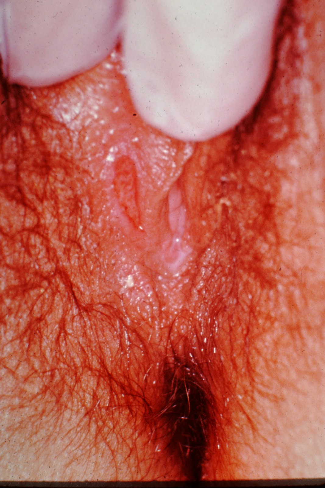
Chancroid

Chancroid is an exquisitely painful ulcerative infection which is sexually transmitted.  It has an incubation period of a few days to two weeks, starts out as a somewhat painful papule which then turns into an ulceration (or ulcerations) which is sharply defined, has ragged borders, bleeds if traumatized and has painful lymphadenopathy.

Diagnostically, the patient would have tender lymphadenopathy, which sometimes suppurates (HSV would NEVER do this), the serology and darkfield is negative for  T Pallidum, the Tzanck Smear is negative for HSV.  Primary HSV tends to have multiple grouped ulcerations as well, but a careful Tzanck will sniff out the right answer.

Thus ends our "Summer of Love" series of STDs.  I hope to have more mundane, less disgusting pics for you in the coming week.  Happy Fourth, everyone!


In more specific terms, the CDC's standard clinical definition for a probable case of chancroid includes all of the following:
  • Patient has one or more painful genital ulcers. The combination of a painful ulcer with tender adenopathy is suggestive of chancroid; the presence of suppurative adenopathy is almost pathognomonic.
  • No evidence of Treponema pallidum is indicated by dark-field examination of ulcer or by a serologic test for Syphilis performed at least 7 days after the onset of ulcer.
About half of infected men have only a single ulcer. Women frequently have four or more ulcers, with fewer symptoms. The ulcers appear in specific locations, such as the coronal sulcus of the uncircumcised glans penis in men, or the fourchette and labia minora in women.

[edit]Common locations in women

In women, the most common location for ulcers is the labia majora. "Kissing ulcers" may develop. These are ulcers that occur on opposing surfaces of the labia. Other areas such as the labia minora,perineal area, and inner thighs may also be involved. The most common symptoms in women are dysuria (pain with urination) and dyspareunia (pain with intercourse).
The initial ulcer may be mistaken as a "hard" chancre, the typical sore of primary syphilis, as opposed to the "soft chancre" of chancroid.
Approximately one-third of the infected individuals will develop enlargements of the inguinal lymph nodes, the nodes located in the fold between the leg and the lower abdomen.
Half of those who develop swelling of the inguinal lymph nodes will progress to a point where the nodes rupture through the skin, producing draining abscesses. The swollen lymph nodes and abscesses are often referred to as buboes.

[edit]Causes

Chancroid is a bacterial infection caused by the fastidious Gram-negative streptobacillus Haemophilus ducreyi. It is a disease found primarily in developing countries, most prevalent in low socioeconomic groups, associated with commercial sex workers.
Infection levels are low in the Western world, typically around one case per two million of the population (Canada, France, Australia, UK and US).[citation needed] Most individuals diagnosed with chancroid have visited countries or areas where the disease is known to occur frequently, although outbreaks have been observed in association with crack cocaine use and prostitution.[citation needed]
Chancroid is a risk factor for contracting HIV, due to their ecological association or shared risk of exposure, and biologically facilitated transmission of one infection by the other.

[edit]Treatment

The CDC recommendation for chancroid is a single oral dose (1 gram) of Azithromycin or a single IM dose of Ceftriaxone or oral Erythromycin for seven days.

[edit]Comparison with syphilis

There are many differences and similarities between the conditions syphilitic chancre and chancroid (reference 1 and 2)
Similarities
  • Both originate as pustules at the site of inoculation, and progress to ulcerated lesions
  • Both lesions are typically 1–2 cm in diameter
  • Both lesions are caused by sexually transmissible organisms
  • Both lesions typically appear on the genitals of infected individuals
  • Both lesions can be present at multiple sites and with multiple lesions
Differences
  • Chancre is a lesion typical of infection with the bacterium that causes syphilis, Treponema pallidum
  • Chancroid is a lesion typical of infection with the bacterium Haemophilus ducreyi
  • Chancres are typically painless, whereas chancroid are typically painful
  • Chancres are typically non-exudative, whereas chancroid typically have a grey or yellow purulent exudate
  • Chancres have a hard (indurated) edge, whereas chancroid have a soft edge
  • Chancres heal spontaneously within three to six weeks, even in the absence of treatment
  • Chancres can occur in the pharynx as well as on the genitals

[edit]