Tuesday, May 22, 2012


Cobblestoning from Cowden's Disease


Cowden's Disease is an autosomal dominant condition with variable penetrance known for a constellation of disease manifestations, all related to abnormal genetic expression, primarily through multiple hamartomas, which lead to its other name, Multiple Hamartoma Syndrome.

The skin findings include trichilemmomas (you should make sure all pts who have trichilemmomas on biopsy do not have Cowden's), oral cobblestoning, skin papillomas, as well as gut, neurologic and, most important, breast lesions and thyroid lesions.  Both can lead to cancer, with female breast cancer in greater than 75% of patients.

Several of you mentioned gingival hyperplasia, and the distingishing difference between this and gingival hyperplasia is that you will see overgrowth of the gums, particularly at the papillae, for hyperplasia, whereas in the cobblestoned mucosa you merely see papillomatosis without the overgrowth.

For those of you who nailed this: nice work.  It's rare, it's important and this is a pretty good example from which to learn.


[edit]

Monday, May 21, 2012


Linear IgA Bullous Dermatosis

Don't worry, the cheese isn't slipping off my cracker, but I did want to run this one as a test to see how the retention of the material previously submitted has gone.  This is apic of the same patient I previously submitted in mid-January and I will include the discussion below, for a refresher.  For the record, the vast majority of you nailed this one, and most noted the "string of pearls".

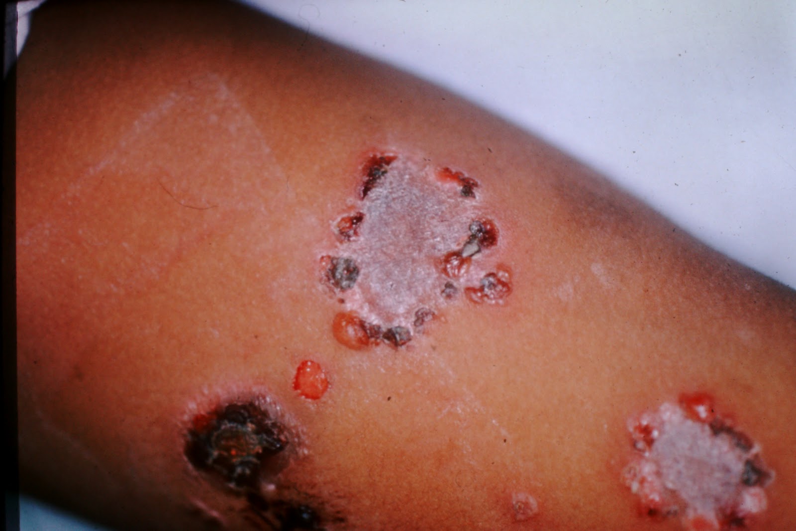
Here is the previous writeup:  Wow!  What a tough question to start out the week!  I want to point out some clinical features as well as some gamesmanship pointers to help you with questions like this.  When looking at an unknown, wither in clinic or in conference, the first question I ask myself is: what is the primary lesion?  That will dictate every other thought I devote to the problem.  In this particular case, I see blisters in a ring around thrashed epidermis.  So, the primary lesion is a blister.  Second, do the blisters look fragile or full thickness?  In my mind, these look to be full thickness, which immediately rules out pemphigus and its variants.  Location?  Looks like the inner forearm, which knocks out PCT and pseudoporphyria, since both of those are predominantly on sun damaged skin.  So, now we can choose among full thickness blisters like LN2, cantharidin, second degree burn, BP. BP variants, bullous LE, or Linear IgA Bullous Dermatosis.  A relatively short, manageable list.  It looks way too patterned for BP, BLE, or LN2.  And so now we are down to cantharidin, burn or LIGA.  Cantharadin is almost always applied only to warts, and the inner forearm is unlikely to develop warts, at least to any significant degree, I told you this is a secondary disease (the patient was treated for something else) and so that rules out burns.  What does that leave?  Linear IgA.  Not only that, but this patient displays the classic "string of pearls" look of blisters in an arc or a ring, which is the most common description of LIGA.  

Why not include DH in this DDX?  Because the blisters are intact in many cases, which is nearly impossible in a DH patient because they are scratching them to beat the band.  Not only that, but they usually look like ant bites.  LOTS of PMNs are recruited into the lesions of DH.  Additionally, this is an unusual location for DH.

What about impetigo?  Well, I gave you the clue that this was a secondary process (see above).  Also, no crusting, although the crusting admittedly isn't always there.

What causes LIGA?  Almost always, it's Vancomycin.  That is always the default answer for LIGA.  Other drugs can do it, but always put your money on Vanc.

What is Vanc used for?  Pseudomembranous colitis caused by C. difficile, and out of control MRSA in all its protean manifestations (joints, endocarditis, etc.).  I would have given points for either answer.

Now, for testmanship:  By the way the Q was written, you should assume that the disease the patient was being treated for was something other than what we see.  Additionally, the "etiologic agent" part of the Q implies that something that was part of the treatment has something to do with the disease we see.  Logically, that would have to be a drug of some sort.  Finally, as mentioned above, a lot of answers are eliminated simply by location.  

As a final point, when I taught immunobullous diseases at Wilford Hall (the Air Force hospital in San Antonio where I did my residency) I used to joke that it took three hands to do a biopsy on a patient like this:  One to hold the punch, one to steady the patient and one to push the biopsying hand away from the primary lesion.  The point is, when doing an immunofluorescence biopsy, make sure you biopsy well away from the blisters, preferably at least 1-2 cm away.  Otherwise, the inflammation will eat up the IgA you want to display on IF, and as a result you can have a false negative IF.

Friday, May 18, 2012


Pseudocyst of the Auricle

The entity known as pseudocyst of the auricle is a relatively uncommon defect in which the cartilaginous plates that make up the auricle (the anterior and posterior leaf) do not join completely.  Sometimes they are traumatically separated and form a seroma-like entity, other times it is more or less a congenital malformation.  It is identified by the relatively smooth swelling, nearly always abutting the helix, but not really involving the helix, leaving it well defined.  It looks deep, much like a lipoma looks deep, as opposed to the other major entity which causes auricular swelling, hemoperichondrium, which leads to cauliflower ear.  Since cauliflower ear is much more superficial, it has the appearance more like a cyst would look on the skin.  Other clues:  cauliflower ear is much more irregular, usually multilobulated, and it usually involves the helix to some degree.  

The other ddx to consider is Stahl's Ear, which is a cartilaginous abnormality of structure which causes the scapha to be flipped forward due to an abnormal additional crus of the anthelix.  If you want extra credit this weekend, forward this link to someone you love who is younger than 18, and then tell me who (in general terms, e.g. your nephew or neighbor kid or whatever) you forwarded it to:   http://dcmf.ca/   .

Thursday, May 17, 2012


Cutis Verticis Gyrata

Cutis verticis gyrata is a condition of the scalp, seen most commonly in African American males, which causes furrowing and ridging of the scalp.  It can be primary, or secondary to a zillion different conditions, most importantly insulin resistant conditions and chronic pulmonary disease.  .  The giveaway on this guy is the clear ridge/furrow pattern to the scalp, something recognizable in spite of the overexposure.  It was described by Alibert, and there's an extra bonus point for ya if you can tell me the hospital with which Alibert was associated.


Wednesday, May 16, 2012


Tendinous xanthomas

Tendinous xanthomas are a deposition of lipids into the macrophages of the skin overlying tendons or ligaments.  Most commonly, you will see these on the hands, feet and overlying the Achilles tendon, but they can also show up in other locations such as the elbows, knees, etc.  They are associated with sky-high lipids, and are commonly considered to be a cutaneous manifestation of internal disease.  How to tell these aren't gouty tophi?  They are too specifically associated with the tendons and joints, especially on the dorsal hands.  How about calcinosis cutis?  Too widely distributed, once again too specifically over the tendons to be calcinosis cutis.  So, everyone understood these were depositions of a substance not native to the skin, but only one of you (Gwen) nailed it.    

Tuesday, May 15, 2012


Coagulopathy

Falstaff, one of Shakespeare's best characters, said in Henry IV "Discretion is the better part of valor", and nothing illustrates that quote better than this picture.  Operating on any patient with a coagulopathy, either iatrogenic (coumadin, Plavix, Effient, steroids, heparin or its analogs, etc.)  or self-induced (as in aspirin, alcohol, Vitamin E, fish oil, garlic, ginseng, gingko biloba, NSAIDS, etc.) or idiopathic (as in this case of ITP, liver failure, coagulation factor deficiencies, etc.) requires a lot of nerve, especially when most of those problems can be corrected with good ol' science.  

This gentleman was operated on for a BCC on the forehead in spite of platelet dysfunction, and although the technicolor results are dramatic, he did quite well in the post op period.  Had the surgeon known he had ITP, no doubt he would have given him a 6 pack or two of platelets, but to be honest, all of us who operate come in contact with patients who are relatively coagulopathic (if I may coin a word).  I usually have at least half of my patients on thinners of one sort or another, and most of them completely ignore my proscriptions against alcohol before surgery.  

So, what's a surgeon to do?  My best advice is to look carefully at the patients when they are biopsied.  If they bleed profusely, but without an obvious reason (as listed above), OR show evidence of extremely abnormal bruising (I saw one of those guys today) then ordering a CBC with plts, and a PT/PTT and an INR for those who are eating rat poison is a good idea.  If they display a coagulopathy and you can't find an obvious cause, send them for a workup via their PCP.  It will save you a lot of patient animosity, diminution of your street cred with the PCPs and sleepless nights.


If you named any reason for coagulopathy, you got credit for the right answer.

For extra credit, how could you reverse a coumadin overdose?


Tuesday, May 1, 2012


Elephantine Tuberculoid Leprosy

Well, this was a free-for-all, with some players entering more than twenty possibilities, some of which  were completely made up out of whole cloth (Bowchickawowow Dz) and some others of which were entirely credible.  Many of the syndromes named indeed had lymphedema, but were either bilateral, or not in healthy people or had other associated signs that were not present in this guy, who, by the way, came from a French book on derm that I have in my library.  I gave as much latitude as I could to each and every suggestion, but some did not meet the credibility criteria, or would have been such an outlier of the disease so as not to be fairly represented.

Here are the diseases I permitted to score as correct:
1. Leprosy
2. Maffucci's Syndrome
3. Cavernous Lymphangioma
4. Filariasis in all its forms 
5. Podoconiosis (my favorite of these dxes)
6. Kaposi's Sarcoma
7. Neurofibromatosis
8. Proteus Syndrome
9. Klippel-Trenaunay Syndrome
10. Chondrosarcoma (I was REALLY liberal with allowing this one, but enough of you tried it, so I allowed it)
11. Lymphangiosarcoma
12. Arteriovenous Malformation
13. Lymphedema secondary to malignancy
14. Onchocerciasis 

Congrats to Emily for winning by two points, and for being the first two-time winner (having won once last year).  So, everybody enjoy your time off, and I will be back in a couple of weeks to challenge you again.  Emily, you get to choose next month's varietal!