Monday, April 30, 2012


Varicella

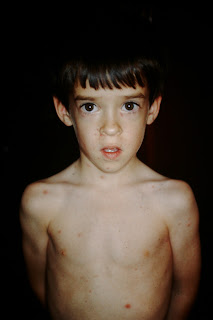
The patient in this picture is my son, Dan, and when he was 5 he came down with chicken pox.  As you can see from the picture, he was afflicted with multiple lesions which were in different stages, showing a bit of a "mouth breather" look (from the oropharyngitis, and he is very young.  Thinking in terms of common things occurring commonly, this would be a "play the odds" kind of question, and if you did (the vast majority of you correctly identified this) you would pick chicken pox.  As you know, varicella is an airborne disease, spread primarily by droplet transmission, and is highly infectious.  Incidentally, this kid just got his first job offer from a think tank in Maryland to do some research in computational linguistics, so that is pretty cool.


Saturday, April 28, 2012


Roswell, New Mexico

Roswell, NM is a strange little town situated in the eastern part of the state, well off the beaten path.  If you found yourself in Roswell, you meant to be there, because there is nothing else around which is worth visiting.  It got its claim to fame because of a strange incident which occurred in late June or early July of 1947, where a UFO was alleged to have crash landed.  

My own (very) tenuous connection with the event is that one of my patients, a local clergyman who was in the seventh grade at the local school at the time, tells me there were some very, very strange things that went on.  He knew the family on whose ranch the UFO was supposedly found, and said those who saw the debris were absolutely convinced the materials were unlike any they had ever seen. According to him, many of the locals saw the crash site. Not only that, there was a family friend who was a nurse who was called in to the clinic where alleged autopsies were performed.  She told several of the townspeople they did autopsies on some kind of creatures she couldn't identify and she seemed very scared.  She disappeared the next day and was never heard from again.  

I went to the museum where they displayed affidavits from career Air Force officers, many of them very high ranking, who swear the craft was not of a material or a technology familiar to them at that time, and some drew the conclusion they were alien craft.

Me?  I don't know.  But, it did make for an interesting afternoon where the museum sold UFO earrings (I bought Cin a pair), they had "My parents went to Roswell, NM and all I got was this lousy tee shirt" and the lightposts were adorned with alien heads.  So, if you are elk hunting on the Mescalero Indian Reservation and you need a break, Roswell is a ticket to a different world.

Thursday, April 26, 2012


Erysipelas

Erysipelas is an infection of the soft tissues, and is generally caused by Group A Beta Hemolytic Streptococcus (GABHS).  It is characterized usually by fever and chills (mediated by the exotoxin secreted by the strep), bright erythema at the site of infection and a peau d'orange (skin of the orange) appearance because the strep toxins cause congestion in the lymphatics, which in turn cause intense edema.  The small dimples are from the hair follicles not being as distensible as the non-adnexal skin.

Usually a result of a small break in the skin, the erysipelas can advance rapidly, and particularly on the face, can lead to sepsis and death.  This guy either gets a bed in the hospital or a spot in the morgue.  I vote for hospitalization.

I gave credit for any of the facial cellulitides, including periorbital cellulitis, and hospitalization/IV abx.
And for those of you who weighed in on the nail question, yes it was Lovibond.  


Wednesday, April 25, 2012


Clubbing Due to Emphysema

Clubbing is classically associated with pulmonary disease, although it can be associated with other conditions, including, of all things, Graves Disease.  The association of vitiligo and clubbing  may make you think of Grave's, but in this particular case it was a red herring.  It was just garden-variety COPD and vitiligo.  Similar appearance is seen in a condition known as hypertrophic pulmonary osteoarthropathy, which can have a destructive arthritic condition as well.  For those of you who have read this far, what is the name of the angle between the nail and the digit, for one point?

Tuesday, April 24, 2012


Toxic Epidermal Necrolysis, healing phase

TEN is a potentially fatal disease that involves the complete sloughing of the epidermis, nearly always as a result of a drug exposure.  It is the most extreme part of the spectrum of erythema multiforme.  Separation of the epidermis happens at the dermal-epidermal level, and the damage can also extend to internal organs, such as the gut and the liver.  The ddx would include a phototoxic reaction, except it is not limited to photoexposed areas; also psoriatic erythroderma would have to be considered.  If you look at the overall process, you get the feeling of skin having just peeled and separated right off the dermis, even at this late stage of healing. These are not the lesions of an immunobullous process, which are more discrete. Also, PRP would have islands of sparing and I would have shown you his palms.  

For those of you who answered the question for extra credit on the treatment of acute paronychia, the answer was indeed incision, drainage and local heat, with possibility of antibiotics (although those are secondary). Good job.

Sunday, April 22, 2012


Acute Paronychia

The big debate s not what it is (paronychia) but whether it is acute or chronic.  Chronic paronychia, as we have seen previously, is a condition that is much like a guerilla war.  Little bit here, little bit there, but no great battles take place.  That is a result of the pathogenic, but not very pathogenic organism which causes chronic paronychia most of the time, and that is Candida sp.  

On the other hand (pun intended) we have acute paronychia.  It is a much more dramatic presentation (YIKES!!), hurts much more and may or may not shoot pus out of the side of the person's finger.  This demands intervention, in a big way.  So, for those of you who have read this far, what IS the intervention for acute paronychia?  One point.  Finally, the name of the organism which most commonly causes acute paronychia is Staph aureus, the golden staph, so named because of its appearance in culture.

Fixed Drug Eruption

This is deceptively difficult. as evidenced by the fact that none of our playas got this one right on the first swing of the bat and you're all pretty damn smart.  But, throw away any preconceptions and just look at the lesion.  

First, it is oval, rather than figurate or angled or irregular in border. Second, look at the border.  It is flat, confluent with the surrounding skin as opposed to raised. The skin is erythematous, but evenly so.  *And most important*, it is the same elevation and texture all the way across the lesion.  If you add all those things up, it can only be an "inside job".  Any external stimulus (contact, spider bite, etc.) would necessarily have a focal point or series of focal points.  

It is well demarcated, which rules out any possibility of necrotizing fasciitis, and to rule out cellulitis, look at how oval it is, the absence of edema, and how demarcated it is.  Can strep be well demarcated and oval?  Yeah, but unlikely, and remember strep causes such a profound local reaction that the body tries to wall it off, causing localized lymphostasis, therefore making it swollen. Erythema Chronicum Migrans is also raised, not flat with the surrounding skin.

Finally, you have the violaceous center.  which cues you in to the FDE.  This is typical for an acute FDE to have a violaceous center.  So, everything I just said should take about 15 seconds to process, and so then the next words out of your mouth are "have you ever had anything like this before?"  


Wednesday, April 18, 2012


Red Scaly Baby Syndrome (Leiner's Disease)

RSB is a tragic entity which is a result of severe immunodeficiency.  Usually C5 deficient, these patients can also be C3 deficient as well as have other immunodeficiencies.  They display severe rashes that look like seb derm, and they often suffer from infections from Gram negatives and yeast.  This disease is usually fatal, but success has been reported with bone marrow transplant.  I gave you all the clues because it is such a good picture of such a rare disease, I thought you should see it and know about it.
Dr. B

Tuesday, April 17, 2012


V1 Herpes Zoster

This was a harder image than I thought it would be.  Several of you missed it, and I don't know why, except that it is a lesion you couldn't just glance at on your iPhone.  To me, when I see certain images, I immediately look to rule out other possibilities on my way to the diagnosis. and in this case I would have eliminated everything else in my ddx because the lesions a) are unilateral and b) extend onto the nose, both at the root and at the sidewall.  Although this is not Hutchinson's Sign, per se, since that is associated with nasal tip involvement, it is nasal involvement and you should refer to ophthalmology, for no other reason than that Morgan and Morgan will smoke you like a cheap cigar if you don't and if the patient gets HZ ophthalmicus.  

For those who do not live in our area, M&M are the slimy ambulance chasers from Orlando who are always on TV.  Of course, the involvement of the nasal tip is also V1 HZV, via the nasociliary nerve (a nerve you should remember all your long lives).  

Monday, April 16, 2012


Numerous melanocytic nevi and lentigines

This question has no specific answer, but was meant to elicit a specific strategy for evaluating and managing patients like this.  The patient has a mix of sunburn lentigines, wildly dysplastic nevi and at least one or two possible melanomas.  So, friends, how do you deal with someone like this who just walks into your office?

The most important part of your game plan is to win this person over, so he trusts you to monitor him, optimally for the rest of his life.  These lesions are guaranteed to change over time, and your job is to pick them off when they start to get too far down the path toward malignancy.  Some of you use dermatoscopes, which are a great tool, but others rely on the naked eye and biopsy those spots which stand out.  

I do not use a dermatoscope.  It is a personal preference, and based on having to remove many melanomas which had been evaluated previously by dermatoscopy and found to be non-malignant.  That being said, I do believe dermatoscopes can at times set your mind at ease about some of the lesions you encounter in the course of a busy day.  So, if you like them, don't let your naked eye be trumped by your dermatoscope, still biopsy the outliers that don't fit with the rest of the lesions. 

The ones I would DEFINITELY biopsy on this guy are the one in the left scapular area and the one on the right upper back with the hyperpigmented poles.  After biopsying those, I would ask him to come back in three months, and keep the close follow ups going until I had biopsied all of the crazy lesions on his back.  I would not feel compelled to biopsy any of his sunburn lentigines.  He should get at the very minimum evaluations every six months.

Since this was such a difficult (can't see the lesions up close) and ambiguous question, I gave you credit for a sharp critical evaluation of the numerous lesions, with a game plan to biopsy the most abnormal ones.


Friday, April 13, 2012


Stephen King's House

On a picture-perfect street in Bangor, Maine sits a pretty Victorian house with the strangest wrought iron fence I've ever seen.  Spiders, webs, bats, it's got 'em all.  Of course, it's all just a joke, but the guy who lives there, Stephen King, is one of America's best selling and most prolific authors.  He owns the market on horror, and despite his creepy subjects and his history of personal demons,  it turns out he is a pretty nice guy.  While we were vacationing in Maine, it seemed that almost everyone we met let us know about the time when they met him, and they all commented on how nice/great/wonderful he was.  He supports innumerable local charities, libraries, Little League teams, and is easily the most beloved man in all of Maine.  Surprisingly, although the gate was closed, the driveway to his house was wide open and his car was parked outside.  I guess he knows nobody there would want to harm him knowingly.   He also happens to be the author of one of the best books I've ever read on how to actually put the pen to the paper called On Writing. His main take home point: avoid adverbs.  He calls them Tom Swifties, after a series of jokes they used to tell back in the day  (40's?,  50's?).  An example:  "Let's play hockey, Tom said puckishly."  Duly noted, carefully.


Darier's Sign

Ferdinand-Jean Darier was the biggest of the big names in French Dermatology, having headed the Department of Derm at HĆ“pital St-Louis in the early 1900's.  His name is everywhere at St-Louis, and he is widely regarded as the father of modern dermatology in the Francophone world, and certainly one of the giants who made our specialty what it is today.  His expertise resided in his ability to observe and describe syndromes and patterns,which in turn led to disease descriptions.  Darier's disease bears his name, but he also described such widely disparate conditions as DFSP, sarcoidosis and erythema annulare centrifugum.  And, of course, the eponymous Darier's sign of urticaria pigmentosa, which would happen if we rubbed these brown spots pictured here.  Also, he headed the Department when Sabouraud (of Sab-Dex fame), Brocq, Fournier (abscess) and Besnier were hanging around at St-Louis.  They were the New York Yankees of dermatology, the Big Five, as it were.  The Big Five (Le Grand Cinq) did not include Baron Dupuytren, another famous French docteur who was universally reviled as "first among surgeons, last among men", which happens to be my favorite insult of all time.  He was at HĆ“tel Dieu.

I gave you credit if you named the sign or the condition.

Thursday, April 12, 2012


Trichotillomania


What’s the First Rule of Dermatology?  Hint:  It's not the same as the First Rule of Fight Club.  The First Rule of Dermatology is this: Don’t believe your ears, believe your eyes.  Trichotillomania is the perfect example of a condition where the history misleads you.  Many, if not most trichotillomaniacs will tell you their scalp, eyelashes, eyebrows, nostril or pubic hair (yes, pubic hair) itches or burns, and that’s why they pull, tug, or scratch these spots.  Really, though, it is the same setup as lichen simplex chronicus.  The chronic trauma makes them itch or burn, then they really go to work.  Many of them will not recognize they are the problem and will deny the deed when pressed.  Always believe your eyes.  The clue in this case is a patient with a condition which does not fit any normal inflammatory pattern.  There is regrowth displayed in the middle (where the TTM started) that is too short for the patient to get a good grip on, but centripetally away from the center there is further alopecia.  Be prepared for an argument when you make the diagnosis, either from the patient or the omnipresent mother, who says “I never see Jessica doing anything like that…”   Uh huh.


Wednesday, April 11, 2012


Two Foot One Hand Disease

Well, dermatologic velociraptors that you are, you all savaged this question pretty ruthlessly.  As you all said, it is two foot- one hand disease, which is caused by (most commonly) Trichophyton rubrum.  Reward yourselves with a different take on the Beatles’ classic “I wanna hold your hand”.  Although you maybe ought to hold off on holding this guy's hand.

Tuesday, April 10, 2012


Pseudoporphyria

Pseudoporphyria is a blistering eruption,usually of the dorsal hands, which is triggered by an exogenous medication.  The most commonly implicated of the meds are the NSAIDs, although a whole host of others have been implicated- even aspirin!

The presentation is a blistering eruption on the dorsal hands, and the ddx is EBA, PCT and PseudoPCT.  Careful history will rule out PCT, and if there is a history of NSAID use in combination with sun exposure, you've got your culprit.

Ten days into April, we've got a three way tie for first, with a bunch of you one point behind.  Keep it up!

Monday, April 9, 2012


Penile Scabies

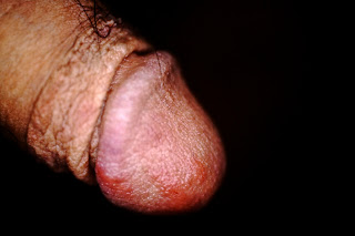
There are lots of different diseases which can pop up on the penis, many of which can be obscure as far as the dx.  Psoriasis, lichen planus, Zoon's balanitis, candida, Lichen sclerosus et atrophicus, allergic contact dermatitis, herpes, all the nasty STDs (chancres, chancroid, etc.) and scabies; but precious few of them itch.  In fact, as a predominant symptom, Pso, LP, Zoon's, Candida, HSV, Chancroid and often LS&A burn or hurt as a presenting symptom.  ACD and scabies: they itch. As a primary symptom, scabies particularly will drive the patients crazy.  As one of our playas said:  "Itchy red papules on the penis are scabies until proven otherwise."  'Nuff said.  Same playa pointed out his favorite spot to look for eggs or mites is underneath the fingernails.  That's a nifty trick for all of you from Redding, California.  Nice job, Brian.


Friday, April 6, 2012


Digital Mucous Cyst

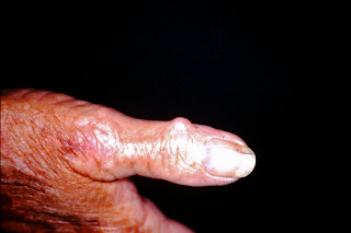
Digital Mucous Cysts are really outpouchings of the synovial joint space of the distal interphalangeal joint.  They are incredibly common in the older age groups, are almost always associated with DIPJ osteoarthritis, and are, of course, benign. They often cause a groove or depression in the nail plate as a result of pressure on the nail matrix, and they are usually asymptomatic.  They are largely of cosmetic consequence only.  At MIMA, if they require treatment, we send them to a hand surgeon.
Many of you picked a synonym for DMCs, and they are allowed.  Happy weekend, Happy Passover and Happy Easter, everyone!

Thursday, April 5, 2012


Positive KOH

KOH exams are one of the fundamental skills we are must possess to be good at taking care of peoples's skin.  The trick to a good KOH exam is to get adequate scale, ensure the patient has not had antifungals prior to the exam, preparing the slide properly and knowing what a positive exam looks like.  

When scraping for a KOH, get far more scale than you think youneed.  Try to get it from inflamed and noninflamed areas, and after scrpaing the scale onto the lside, drop the KOH solution (we use Swarz-Lamkins) onto the slide, and then lightly heat the slide until the small bubbles underneath the cover slip expand, but before the fluid boils.

After slide prep, expect the slide to be readable: no thick scale on the slide, good stain, all the carbon suet removed from the slide.  Examine the slide by using a quick up and down motion of the objective, it will help you identify which are real hyphal elements and which are just cell borders.

This slide is very positive, as demonstrated by the parallel margins of the cell walls of the hyphal elements, the septations and the branches coming off the main hyphae.


Wednesday, April 4, 2012


Microcystic Adnexal Carcinoma

Rather than belabor the relationship between infiltrative BCCs and MACs, I will instead list what is the reasonable differential diagnosis.  I did not count off for wrong answers, but did give credit for right ones. Here's the DDx as accepted by most dermatologists:  Scar, MAC, Infiltrative BCC, Desmoplastic trichoepithelioma, post-LN2 hypopigmentation (although I've never seen it look like this) and vitiligo (never seen it look like this, either.  For those of you who put regressed MM, I will grant that, although it is entirely atypical for that, but I did not give credit for amelanotic MM, because they are pink or red. I really want to make the point that any scar that is irregular or round on the face demands an explanation, and if no history of trauma exists, then biopsy is indicated.  

No infectious causes would be this discrete, and except as above, no inflammatory processes would be, either.

Tuesday, April 3, 2012


Infiltrative Basal Cell Carcinoma

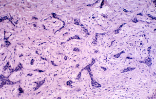
Infiltrative basal cell carcinomas are tough.  They can be small or overwhelmingly large, and often clinical examination doesn't tell just how big they are.  They are best thought of as worm holes down into the dermis, and they look like our image when cut en face for viewing under the microscope.  Normal basal cells have large balls of tumor (which look like little beads when we biopsy them) and usually are surrounded by mucin (which explains why they are sticky and want to adhere to our razor blade or scalpel when we shave biopsy them). 


Think of nodular basal cells as cauliflower.  When you thin slice cauliflower, the slice looks like individual balls of material, but in actuality they were attached to the main stalk of the cauliflower.  Infiltrative basal cells, on the other hand, extend as worm holes down into the tissue, and the way to think of them in 3-D is as if you were thin slicing a mass of cold spaghetti.  if you looked at only the thin slice, it would look almost exactly like this slide: some of the strands would be viewed straight on, others are cut at an angle so as to give the appearance of little spikes and strands of tissue.  

Why bother with this?  Because once you get the "zen" of this tumor, you will be able to understand how to defeat it.  The best way, by far, is by Mohs surgery.  We can track down all the individual strands and tentacles that extend form the main body of the tumor, and that will give you the highest cure rate.  But, you say, I don't do Mohs!  Well, you can either take a wide margin around what you see as the edge of the tumor, or you can use a little trick:  Stretch the skin, and look for blanching,  Since the tumor is a space-occupying lesion in the dermis, it displaces blood vessels and so when you stretch the skin around the tumor, it blanches out the skin where the tumor is.  That is a more accurate way of predicting the margin.  Many of you are doing surgeries, and when you are approaching basals and SCCs it is helpful to define the margins as well as you can.  Along with stretching the skin, also consider curetting the tissue prior to excision for nodular BCCs and SCCs.  It'll cut way down on your positive margin rate.  On infiltratives, that won't work because the strands of tumor are too small for the curette to be able to adequately define.  

The clinical differential diagnoses for these infiltrative tumors are scars, microcystic adnexal carcinomas, a benign growth known as a desmoplastic trichoepithelioma, and the occasional weird large syringoma.  If the infiltrative tumor has increased collagen under the microscope, it is known as "sclerosing", which means "scarring". 

Small clinical tumors can be very large:


Infiltrative BCC post-Mohs

One final point:  nodular basals can be triggered into becoming infiltrative with external trauma, such as consistent scratching, or from partial treatment, especially by using liquid nitrogen on nodular and superficial BCCs.  It is imperative that you biopsy what you don't know, and only freeze those lesions that you KNOW are actinic keratoses.
Monday, April 2, 2012


The Treasury Petra, Jordan


Petra is one of the most amazing places in the world.  It was the center of the Nabatean Kingdom, and was, amazingly, unknown to the outside world until 1812.  The Nabateans were traders whose caravansary worked the area from the Dead Sea down to present day Yemen, at the southern tip of the Arabian Peninsula.  Their power came from these trade routes, which started out from oasis to oasis, but then evolved to a complex network of man-made oases, where cisterns were dug into the rock and thus the annual rains would collect in these cisterns, and then the caravans would use them as watering stations for their camels.  In a way, they made the desert their fortress, because without the knowledge of where these watering holes were, the invaders and bandits were out of luck. They, and they alone, could get the goods from one spot in the desert to another.  They were FedEx before FedEx was FedEx.

Petra was carved from the sandstone of the Jordanian desert in the 300 B.C. era, and was remarkably unnoticeable from the surrounding countryside.  It really amounts to a canyon, with its own water supply (controlled by a sophisticated aqueduct system) and inhospitable surroundings which made invaders more reluctant to pick a fight.  Out of this sandstone canyon were carved spectacular buildings, most of which had Greek-inspired architecture, and the most spectacular of these is The Treasury, which is, as one of our playas pointed out, known as Al Khazneh in Arabic.  There are lots of yarns about why it was called the Treasury, but I'm pretty sure the real reason why is that the Swiss archeologist who discovered it probably labelled it according to his own sensibilities. (Hey, he was Swiss; the Swiss really, really love their banks, and so you see where I'm going with this...)

One of the Indiana Jones movies was shot there, as another playa pointed out, and the area outside the actual historical site is replete with Indiana Jones souvenir shops.  You can take camel rides, but the rides are sort of like the pony rides we used to take as a kid, with the actual Jordanian camel driver leading your camel past all the historic sites, before they drop you off at their cousin's souvenir shop.  They are pushy, but in a roguish, fun sort of way.  Their kids can work a crowd for money better than Barack Obama, and the mothers usually sit in the shadows, scowling at the western women who are immodestly dressed.  Culture meets commerce.  

Of course, the dromedary camel is the ship of the desert, and although they have a face only a mother could love, they are pretty cool.  They take their time going where they are going, grumble loudly if prodded, and act up for no apparent reason.  Instead of calling them the ships of the desert, it's more like "teenagers of the desert".  

Well, my brain has had a break, so it's back to derm maƱana, but many of you hoovered up two points on an interesting little side trip on our road to knowledge.